A Melting Method for RNA Extraction from the Mucosal Membrane of the Mouse Middle Ear
- Affiliations
-
- 1Department of Otorhinolaryngology, Yonsei University College of Medicine, Seoul, Korea. jychoi@yuhs.ac
- KMID: 2070030
- DOI: http://doi.org/10.3349/ymj.2015.56.2.497
Abstract
- PURPOSE
There is much confusion surrounding the methods of RNA extraction from the middle ear mucosa of mice. In this study, we worked to develop a "melting method," which is faster, purer, and more reliable than other methods in common use.
MATERIALS AND METHODS
Thirty-two ears were used for this study. Light microscopy with hematoxylin-eosin staining of the bullae, scanning electron microscopy (SEM), spectrophotometer analysis, and reverse transcription polymerase chain reaction were performed before and after melting the half lateral bullae, which were detached from the temporal bone by using a lateral retroauricular approach.
RESULTS
Each resected half bulla contained a well distributed mucosal membrane. After a TRIzol melting duration of 10-30 minutes, only mucosal marker (MUC5AC) was expressed without bony marker (total osteocalcin). The same results were determined from SEM.
CONCLUSION
This melting method, compared with stripping and irrigation methods, is effective and offers an easier, more robust approach to extracting RNA from the middle ear mucosal membranes of mice.
Keyword
MeSH Terms
Figure
-
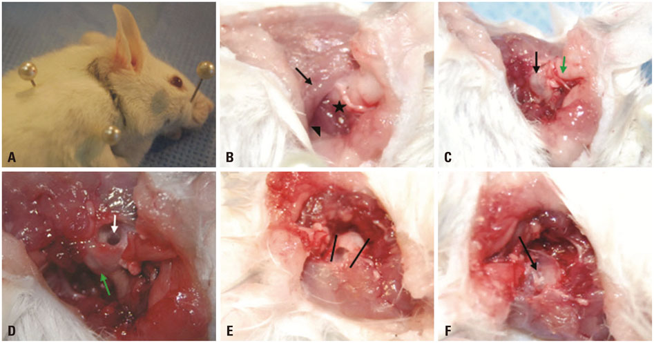
Fig. 1 Lateral postauricular approach to the mouse middle ear (bulla). (A) In each animal, a postauricular incision (black line marker) was made and extended ventrally to the rostral neck skin. The subcutaneous connective tissues were separated to expose the deeper structures. (B) The incision was deepened to the underlying temporalis and cervical muscles. Subsequently, the submaxillary gland was carefully dissected out and retracted, working across the sternocleidomastoid muscle (black arrow) and the digastric muscles (arrowhead). The bulla (star) is the posterior insertion of the digastric muscle into the jugular process of the occipital bone. (C) Access to the tympanic bulla was achieved by means of a blunt dissection slightly lateral to the insertion of the digastric muscle. The figure shows the bulla (black arrow) connected to the EAC (green arrow). (D) The EAC was resected with the tympanic membrane using a probe that was inserted into the EAC and bulged toward the dissection. The previously exposed bulla (green arrow) is inferior to the resected tympanic membrane, showing the middle ear cavity inside (white arrow). (E) Looking through the hole of the bulla upside the mouse, the half-exposed clean bulla (black lines) was resected with sharp micro-scissors, leaving no fragments. (F) After the resection, the medial wall of the bulla, including the promontory (black arrow), remained. EAC, external auditory canal.
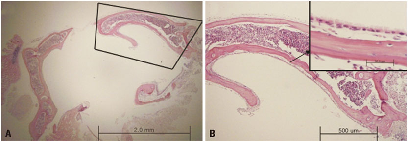
Fig. 2 Light micrographs of the middle ear mucosa in the whole bulla. (A) Inside the bulla bone, ciliated mucosal membranes were evenly distributed. A lateral approach exposed the half lateral bulla (black box) (×10). (B) The resected lateral bulla had securely attached mucosal membranes (×20, ×400).
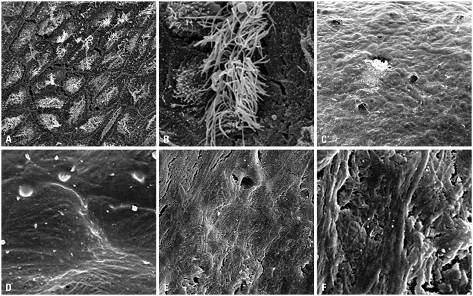
Fig. 3 Scanning electron micrographs of the middle ear mucosa in the resected bulla. (A) Before melting, the picture shows ciliated mucosal cells regularly distributed on the surface of bony bulla (×1000). (B) A bundle of cilia on the hexagonal cell (×10000). (C and D) After melting the membrane into the TRIzol solution for 10 minutes, it showed uneven surfaces and no ciliated cells (×1000, ×10000). There were very few mucosal cells on the surface, and it melted almost all mucosal cells of the bulla, not exposing bony surfaces. (E and F) After stripping, the bony surface was exposed with some bony scratching (×1000, ×10000).
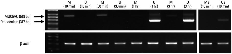
Fig. 4 Expression of mucosal marker (MUC5AC) and bony marker (osteocalcin) in mouse middle ear epithelium by RT-PCR; 518-bp and 371-bp fragments amplified on the RT-PCR probed by MUC5AC and total osteocalcin primers. MUC5AC was detected in the cells within 1 hour, while MUC5AC and osteocalcin were simultaneously detected over a period of 1 hour. RT-PCR, reverse transcription-polymerase chain reaction; M, MUC5AC primer in the melting method; O, osteocalcin primer in the melting method; Ms, MUC5AC primer in the stripping method; Os, osteocalcin primer in the stripping method.
Reference
-
1. Wysocki J. Topographical anatomy and measurements of selected parameters of the rat temporal bone. Folia Morphol (Warsz). 2008; 67:111–119.2. Sichel JY, Plotnik M, Cherny L, Sohmer H, Elidan J. Surgical anatomy of the ear of the fat sand rat. J Otolaryngol. 1999; 28:217–222.3. Dogru S, Haholu A, Gungor A, Kucukodaci Z, Cincik H, Ozdemir T, et al. Histologic analysis of the effects of three different support materials within rat middle ear. Otolaryngol Head Neck Surg. 2009; 140:177–182.
Article4. Nell MJ, Grote JJ. Structural changes in the rat middle ear mucosa due to endotoxin and eustachian tube obstruction. Eur Arch Otorhinolaryngol. 1999; 256:167–172.
Article5. Fleige S, Pfaffl MW. RNA integrity and the effect on the real-time qRT-PCR performance. Mol Aspects Med. 2006; 27:126–139.
Article6. Desbois C, Hogue DA, Karsenty G. The mouse osteocalcin gene cluster contains three genes with two separate spatial and temporal patterns of expression. J Biol Chem. 1994; 269:1183–1190.
Article7. Kerschner JE, Li J, Tsushiya K, Khampang P. Mucin gene expression and mouse middle ear epithelium. Int J Pediatr Otorhinolaryngol. 2010; 74:864–868.
Article8. Judkins RF, Li H. Surgical anatomy of the rat middle ear. Otolaryngol Head Neck Surg. 1997; 117:438–447.
Article9. Cayé-Thomasen P, Hermansson A, Tos M, Prellner K. Increased secretory capacity of the middle ear mucosa after acute otitis media caused by Haemophilus influenzae type B. Otolaryngol Head Neck Surg. 1997; 117(3 Pt 1):263–267.10. Palacios SD, Pak K, Rivkin AZ, Kayali AG, Austen D, Aletsee C, et al. Role of p38 mitogen-activated protein kinase in middle ear mucosa hyperplasia during bacterial otitis media. Infect Immun. 2004; 72:4662–4667.
Article11. Chen YP, Tong HH, James , Demaria TF. Detection of mucin gene expression in normal rat middle ear mucosa by reverse transcriptase-polymerase chain reaction. Acta Otolaryngol. 2001; 121:45–51.
Article12. Furukawa M, Ebmeyer J, Pak K, Austin DA, Melhus A, Webster NJ, et al. Jun N-terminal protein kinase enhances middle ear mucosal proliferation during bacterial otitis media. Infect Immun. 2007; 75:2562–2571.
Article13. Pinilla M, Ramírez-Camacho R, Jorge E, Trinidad A, Vergara J. Ventral approach to the rat middle ear for otologic research. Otolaryngol Head Neck Surg. 2001; 124:515–517.
Article
- Full Text Links
-
- Actions
-
Cited
- CITED
-
- Close
- Share
-
- Similar articles
-
- Expression of beta Defensins in the Human Middle Ear Mucosa
- The Effects of Antibiotics and Steroid on the Middle Ear Mucosa in the Rats with Experimental Acute Otitis Media
- Cholesterol Granuloma without Tympanic Membrane Perforation
- Tympanic Membrane Perforation Due to Metal Spark in a Welder
- A Case of Intratympanic Membrane Congenital Cholesteatoma
