Cancer Stem Cells in Primary Liver Cancers: Pathological Concepts and Imaging Findings
- Affiliations
-
- 1Department of Radiology, Seoul National University Hospital, Seoul 110-744, Korea. jmsh@snu.ac.kr
- 2Department of Pathology, Seoul National University Bundang Hospital, Seongnam 463-707, Korea.
- KMID: 2069984
- DOI: http://doi.org/10.3348/kjr.2015.16.1.50
Abstract
- There is accumulating evidence that cancer stem cells (CSCs) play an integral role in the initiation of hepatocarcinogenesis and the maintaining of tumor growth. Liver CSCs derived from hepatic stem/progenitor cells have the potential to differentiate into either hepatocytes or cholangiocytes. Primary liver cancers originating from CSCs constitute a heterogeneous histopathologic spectrum, including hepatocellular carcinoma, combined hepatocellular-cholangiocarcinoma, and intrahepatic cholangiocarcinoma with various radiologic manifestations. In this article, we reviewed the recent concepts of CSCs in the development of primary liver cancers, focusing on their pathological and radiological findings. Awareness of the pathological concepts and imaging findings of primary liver cancers with features of CSCs is critical for accurate diagnosis, prediction of outcome, and appropriate treatment options for patients.
Keyword
MeSH Terms
-
Bile Duct Neoplasms/pathology/radiography
Bile Ducts, Intrahepatic/pathology/radiography
Carcinoma, Hepatocellular/pathology/radiography
Cholangiocarcinoma/pathology/radiography
Humans
Liver Neoplasms/*pathology/radiography
Magnetic Resonance Imaging
Neoplastic Stem Cells/*pathology/radiography
Tomography, X-Ray Computed
Figure
-
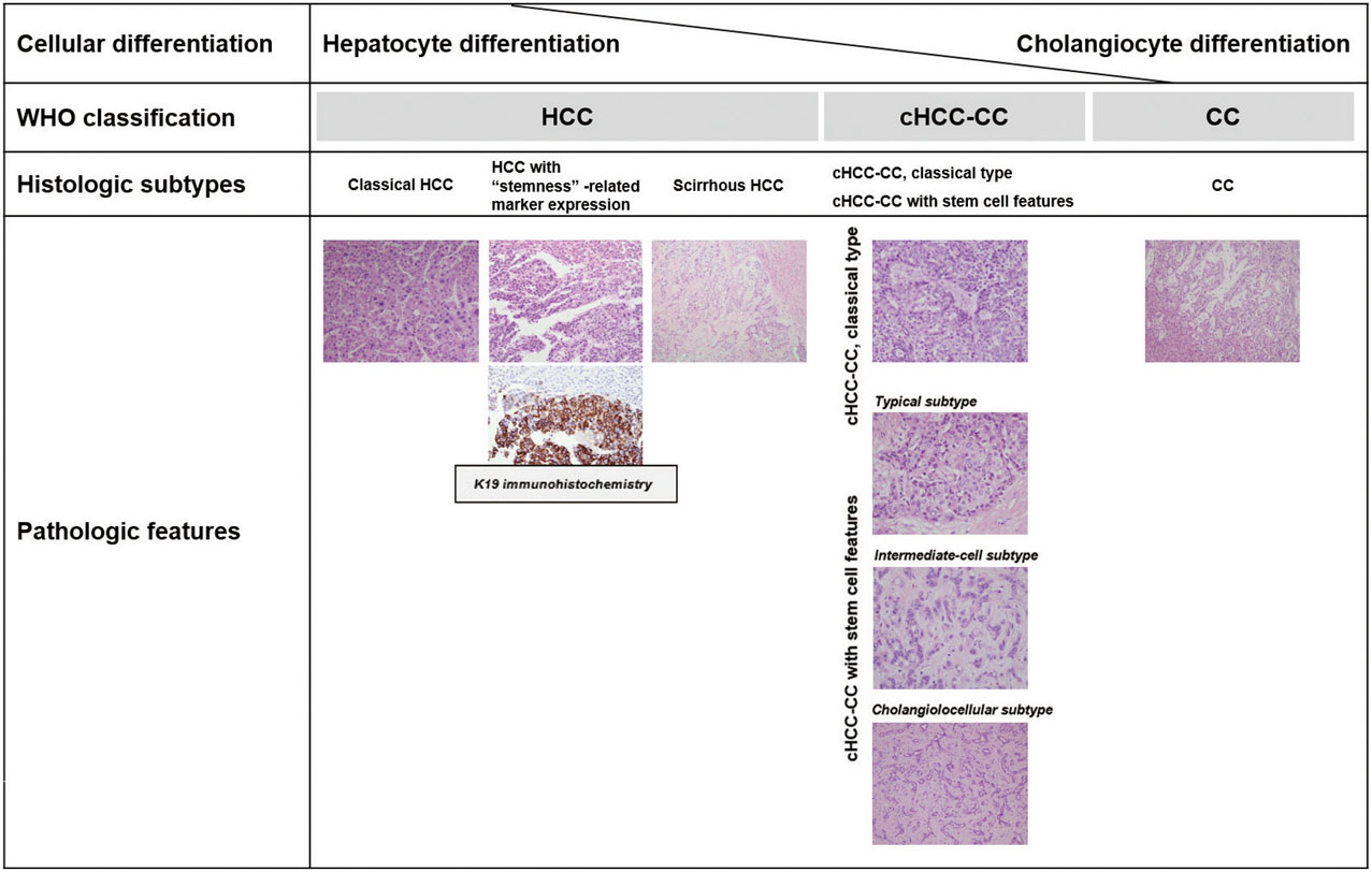
Fig. 1 Spectrum of primary liver cancers according to cellular differentiation. CC = cholangiocarcinoma, cHCC-CC = combined hepatocellular-cholangiocarcinoma, HCC = hepatocellular carcinoma, WHO = World Health Organization
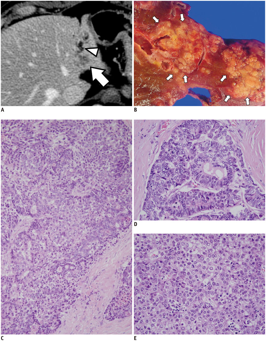
Fig. 2 Pathologically confirmed classical type of cHCC-CC. A. Portal phase CT image showing ill-defined mass (arrow) in left lobe of liver and dilated intrahepatic duct (arrowhead). B. On gross specimen showing ill-defined infiltrative solid mass (arrows). C-E. Microscopic findings revealing mixture of glandular (CC) and hepatocytic (HCC) differentiation. CC component (D) and HCC component (E) are shown in greater detail on right panel (H&E stain, × 100 (C), × 200 (D, E)). CC = cholangiocarcinoma, cHCC-CC = combined hepatocellular-cholangiocarcinoma, HCC = hepatocellular carcinoma
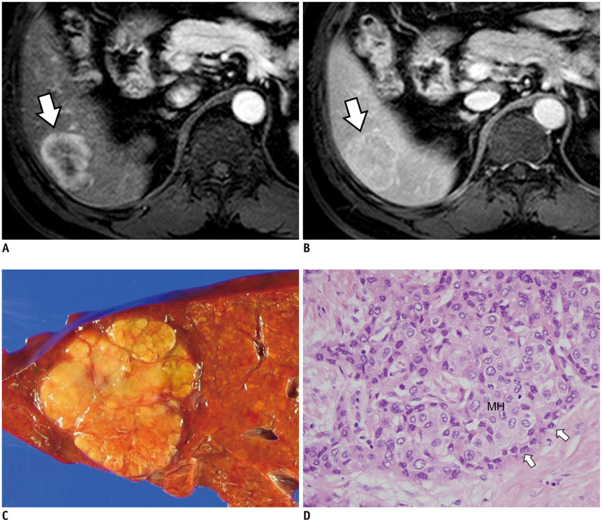
Fig. 3 cHCC-CC with stem cell features of typical stem cell subtype in 58-year-old male patient with chronic hepatitis B with moderate elevation of serum AFP level (154 IU/mL). A. MR image on arterial phase showing 4 cm tumor (arrow) with peripheral enhancement in segment 6 of liver. B. Hepatic mass (arrow) showing centripetal enhancement on portal phase image. C. Photograph of gross specimen showing lobulated solid yellowish-white mass. D. On microscopic examination, tumor is composed of sheets and islands of polygonal mature-looking hepatocyte-like tumor cells (MH) with eosinophilic cytoplasm surrounded by peripheral rim of smaller tumor cells with high nuclear:cytoplasmic ratio resembling hepatic stem/progenitor cells (arrows) (H&E stain, × 400). AFP = alpha-fetoprotein, cHCC-CC = combined hepatocellular-cholangiocarcinoma
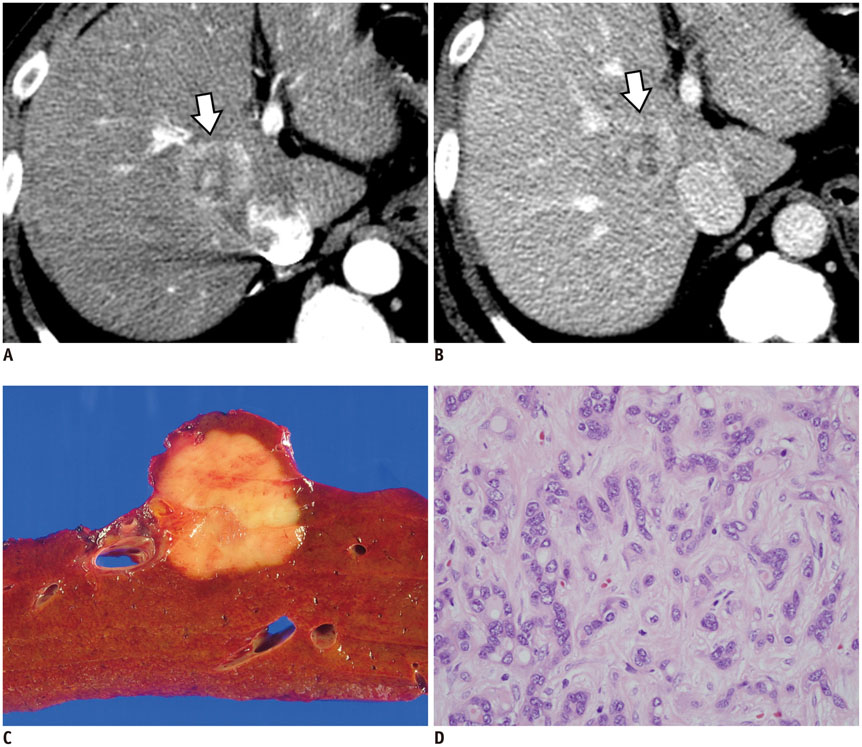
Fig. 4 Combined hepatocellular-cholangiocarcinoma with stem cell features of intermediate cell subtype in 53-year-old male patient. A. Arterial phase CT scan demonstrating hyperenhancing mass (arrow) in segment 8 of liver. B. Portal phase CT scan showing prolonged enhancement of mass without washout (arrow). C. On gross examination, homogeneous yellowish-white solid lobulated mass is seen. D. Trabeculae and cords of relatively monomorphic population of tumor cells with high nuclear:cytoplasmic ratio in background of fibrotic stroma are seen (H&E stain, × 400).

Fig. 5 Combined hepatocellular-cholangiocarcinoma with stem cell features of cholangiolocellular subtype in 67-year-old male patient with alcoholic liver cirrhosis. A. Precontrast T1-weighted MRI showing lobulated well-defined mass (arrow) in segment 6 of liver. Note hepatic surface retraction (arrowhead). Arterial (B) and delayed phase images (C) demonstrating gradual enhancement of lateral portion of tumor (arrows). D. Solid greyish-white mass with indistinct margins. E. Tumor is characterized by distinct tubular structures embedded in fibrotic stroma, reminiscent of ductular reactions (H&E stain, × 200).

Fig. 6 Schematic representation of imaging features of HCC, cHCC-CC, and CC. A, C, E. Arterial phase images. B, D, F. Portal and delayed phase images. A, B. HCC in cirrhotic liver shows arterial enhancement and delayed washout. Pseudocapsule shows delayed enhancement. C, D. cHCC-CC in cirrhotic liver shows strong arterial enhancement in peripheral portion of tumor and concentric filling on delayed phase. Note tumor thrombus in portal vein. E, F. CC in noncirrhotic liver shows weak arterial enhancement in peripheral portion of tumor and concentric filling on delayed phase. Note bile duct dilation and hepatic surface retraction. CC = cholangiocarcinoma, cHCC-CC = combined hepatocellular-cholangiocarcinoma, HCC = hepatocellular carcinoma
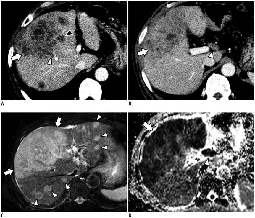
Fig. 7 Pathologically confirmed cHCC-CC with vascular invasion and lymph node metastases in 60-year-old male patient with liver cirrhosis. A, B. Contrast-enhanced CT images of different sections showing large lobulated mass (arrows) with poor enhancement. A. Note tumor thrombus in portal vein branch in segment 8 (white arrowhead) and in middle hepatic vein (black arrowhead). B. Metastatic hepatoduodenal lymph node (arrowhead). C. Fat-suppressed T2-weighted image showing large high signal intensity mass involving segment 4 and right anterior segment of liver (arrows) as well as multiple daughter nodules (arrowheads). D. ADC map showing diffusion restriction of hepatic mass (arrow) and metastatic hepatoduodenal lymph node (arrowhead). ADC = apparent diffusion coefficient, cHCC-CC = combined hepatocellular-cholangiocarcinoma

Fig. 8 Pathologically confirmed combined hepatocellular-cholangiocarcinoma with stem cell features of cholangiolocellular subtype in 58-year-old male patient with chronic hepatitis B. A. Fat-suppressed T2-weighted image revealing lobulated mass (arrow) in right posterior segment of liver with moderately high signal intensity in peripheral portion of mass. On arterial phase (B) and portal phase (C) of gadoxetic acid-enhanced MR T1-weighted images, mass demonstrates strong arterial enhancement in peripheral portion and delayed concentric enhancement without washout (arrows). D. Hepatobiliary phase T1-weighted image showing mass as clear hypointense lesion. Note hepatic surface retraction (arrowhead). E. Solid lobulated on gross examination.
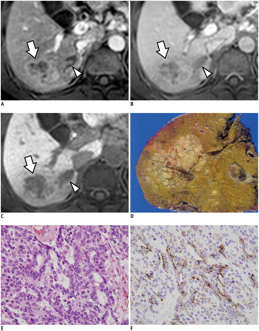
Fig. 9 Hepatocellular carcinoma with "stemness"-marker expression in 48-year-old male patient with liver cirrhosis. A-C. Gadoxetic acid-enhanced MRI showing irregular mass (arrows) in segment 6 of liver with tumor thrombus (arrowheads) in right inferior hepatic vein. Mass shows peripheral enhancement on arterial phase (A) and delayed enhancement (B). C. Hepatobiliary phase showing mass with hypointensity. D. Gross examination reveals infiltrative solid greyish-white mass in liver. E. Tumor cells with eosinophilic cytoplasm arranged in sheets and pseudoglandular structures (H&E stain, × 400). F. Immunohistochemical staining revealing K19-positive cells in tumor (× 400).
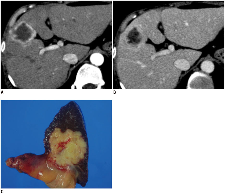
Fig. 10 Scirrhous hepatocellular carcinoma in 50-year-old female patient with liver cirrhosis. A. Arterial phase CT image showing lobulated mass with peripheral enhancement in segment 8 of liver. B. Portal phase CT image demonstrating prolonged centripetal enhancement of tumor. C. Specimen photograph showing lobulated solid yellowish-white mass.
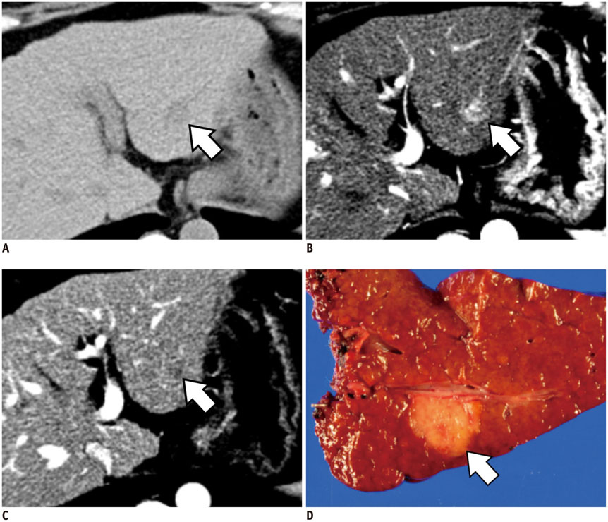
Fig. 11 Intrahepatic CC mimicking HCC in background of chronic liver disease in 63-year-old female patient. A. Precontrast CT image showing small low-attenuated mass (arrow) in segment 2 of liver. Note surface nodularity of liver suggesting liver cirrhosis. B. Arterial phase CT image showing mass with hyperenhancement (arrow). C. Portal phase CT image demonstrating washout of mass (arrow). This dynamic enhancement pattern is suggestive of HCC. D. Specimen photograph revealing lobulated yellowish mass (arrow) which was pathologically confirmed as intrahepatic CC. Note background cirrhotic liver. CC = cholangiocarcinoma, HCC = hepatocellular carcinoma
Cited by 1 articles
-
Atypical Appearance of Hepatocellular Carcinoma and Its Mimickers: How to Solve Challenging Cases Using Gadoxetic Acid-Enhanced Liver Magnetic Resonance Imaging
Jae Hyun Kim, Ijin Joo, Jeong Min Lee
Korean J Radiol. 2019;20(7):1019-1041. doi: 10.3348/kjr.2018.0636.
Reference
-
1. Nguyen LV, Vanner R, Dirks P, Eaves CJ. Cancer stem cells: an evolving concept. Nat Rev Cancer. 2012; 12:133–143.2. Hill RP, Perris R. "Destemming" cancer stem cells. J Natl Cancer Inst. 2007; 99:1435–1440.3. Marquardt JU, Factor VM, Thorgeirsson SS. Epigenetic regulation of cancer stem cells in liver cancer: current concepts and clinical implications. J Hepatol. 2010; 53:568–577.4. Lo RC, Ng IO. Hepatic progenitor cells: their role and functional significance in the new classification of primary liver cancers. Liver Cancer. 2013; 2:84–92.5. Roskams T. Liver stem cells and their implication in hepatocellular and cholangiocarcinoma. Oncogene. 2006; 25:3818–3822.6. Allen RA, Lisa JR. Combined liver cell and bile duct carcinoma. Am J Pathol. 1949; 25:647–655.7. Fowler KJ, Sheybani A, Parker RA 3rd, Doherty S, M Brunt E, Chapman WC, et al. Combined hepatocellular and cholangiocarcinoma (biphenotypic) tumors: imaging features and diagnostic accuracy of contrast-enhanced CT and MRI. AJR Am J Roentgenol. 2013; 201:332–339.8. Libbrecht L. Hepatic progenitor cells in human liver tumor development. World J Gastroenterol. 2006; 12:6261–6265.9. Bota S, Piscaglia F, Marinelli S, Pecorelli A, Terzi E, Bolondi L. Comparison of international guidelines for noninvasive diagnosis of hepatocellular carcinoma. Liver Cancer. 2012; 1:190–200.10. Song DS, Bae SH. Changes of guidelines diagnosing hepatocellular carcinoma during the last ten-year period. Clin Mol Hepatol. 2012; 18:258–267.11. Oliva MR, Saini S. Liver cancer imaging: role of CT, MRI, US and PET. Cancer Imaging. 2004; 4(Spec No A):S42–S46.12. Joo I, Choi BI. New paradigm for management of hepatocellular carcinoma by imaging. Liver Cancer. 2012; 1:94–109.13. Joo I, Lee JM. Imaging bile duct tumors: pathologic concepts, classification, and early tumor detection. Abdom Imaging. 2013; 38:1334–1350.14. Teefey SA, Hildeboldt CC, Dehdashti F, Siegel BA, Peters MG, Heiken JP, et al. Detection of primary hepatic malignancy in liver transplant candidates: prospective comparison of CT, MR imaging, US, and PET. Radiology. 2003; 226:533–542.15. Lee WS, Lee KW, Heo JS, Kim SJ, Choi SH, Kim YI, et al. Comparison of combined hepatocellular and cholangiocarcinoma with hepatocellular carcinoma and intrahepatic cholangiocarcinoma. Surg Today. 2006; 36:892–897.16. Durnez A, Verslype C, Nevens F, Fevery J, Aerts R, Pirenne J, et al. The clinicopathological and prognostic relevance of cytokeratin 7 and 19 expression in hepatocellular carcinoma. A possible progenitor cell origin. Histopathology. 2006; 49:138–151.17. Ikeda H, Harada K, Sato Y, Sasaki M, Yoneda N, Kitamura S, et al. Clinicopathologic significance of combined hepatocellular-cholangiocarcinoma with stem cell subtype components with reference to the expression of putative stem cell markers. Am J Clin Pathol. 2013; 140:329–340.18. Fan L, He F, Liu H, Zhu J, Liu Y, Yin Z, et al. CD133: a potential indicator for differentiation and prognosis of human cholangiocarcinoma. BMC Cancer. 2011; 11:320.19. Cardinale V, Carpino G, Reid L, Gaudio E, Alvaro D. Multiple cells of origin in cholangiocarcinoma underlie biological, epidemiological and clinical heterogeneity. World J Gastrointest Oncol. 2012; 4:94–102.20. Komuta M, Govaere O, Vandecaveye V, Akiba J, Van Steenbergen W, Verslype C, et al. Histological diversity in cholangiocellular carcinoma reflects the different cholangiocyte phenotypes. Hepatology. 2012; 55:1876–1888.21. Ijichi H, Shirabe K, Taketomi A, Yoshizumi T, Ikegami T, Mano Y, et al. Clinical usefulness of (18) F-fluorodeoxyglucose positron emission tomography/computed tomography for patients with primary liver cancer with special reference to rare histological types, hepatocellular carcinoma with sarcomatous change and combined hepatocellular and cholangiocarcinoma. Hepatol Res. 2013; 43:481–487.22. Ji J, Wang XW. Clinical implications of cancer stem cell biology in hepatocellular carcinoma. Semin Oncol. 2012; 39:461–472.23. Yamashita T, Wang XW. Cancer stem cells in the development of liver cancer. J Clin Invest. 2013; 123:1911–1918.24. Andersen JB, Loi R, Perra A, Factor VM, Ledda-Columbano GM, Columbano A, et al. Progenitor-derived hepatocellular carcinoma model in the rat. Hepatology. 2010; 51:1401–1409.25. Wong DJ, Liu H, Ridky TW, Cassarino D, Segal E, Chang HY. Module map of stem cell genes guides creation of epithelial cancer stem cells. Cell Stem Cell. 2008; 2:333–344.26. Yang XR, Xu Y, Yu B, Zhou J, Qiu SJ, Shi GM, et al. High expression levels of putative hepatic stem/progenitor cell biomarkers related to tumour angiogenesis and poor prognosis of hepatocellular carcinoma. Gut. 2010; 59:953–962.27. Chiba T, Kita K, Zheng YW, Yokosuka O, Saisho H, Iwama A, et al. Side population purified from hepatocellular carcinoma cells harbors cancer stem cell-like properties. Hepatology. 2006; 44:240–251.28. Theise ND, Nakashima O, Park YN, Nakanuma Y. Combined hepatocellular-cholangiocarcinoma. In : Bosman FT, Carneiro F, Hruban RH, Theise ND, editors. WHO classification of tumours of the digestive system. 4th ed. Lyon: IARC;2010. p. 225–227.29. Lin G, Toh CH, Wu RC, Ko SF, Ng SH, Chou WC, et al. Combined hepatocellular cholangiocarcinoma: prognostic factors investigated by computed tomography/magnetic resonance imaging. Int J Clin Pract. 2008; 62:1199–1205.30. Goodman ZD, Ishak KG, Langloss JM, Sesterhenn IA, Rabin L. Combined hepatocellular-cholangiocarcinoma. A histologic and immunohistochemical study. Cancer. 1985; 55:124–135.31. Kim H, Park C, Han KH, Choi J, Kim YB, Kim JK, et al. Primary liver carcinoma of intermediate (hepatocyte-cholangiocyte) phenotype. J Hepatol. 2004; 40:298–304.32. Terada T. Combined hepatocellular-cholangiocarcinoma with stem cell features, ductal plate malformation subtype: a case report and proposal of a new subtype. Int J Clin Exp Pathol. 2013; 6:737–748.33. Theise ND, Yao JL, Harada K, Hytiroglou P, Portmann B, Thung SN, et al. Hepatic 'stem cell' malignancies in adults: four cases. Histopathology. 2003; 43:263–271.34. Komuta M, Spee B, Vander Borght S, De Vos R, Verslype C, Aerts R, et al. Clinicopathological study on cholangiolocellular carcinoma suggesting hepatic progenitor cell origin. Hepatology. 2008; 47:1544–1556.35. Shiota K, Taguchi J, Nakashima O, Nakashima M, Kojiro M. Clinicopathologic study on cholangiolocellular carcinoma. Oncol Rep. 2001; 8:263–268.36. Shetty AS, Fowler KJ, Brunt EM, Agarwal S, Narra VR, Menias CO. Combined hepatocellular-cholangiocarcinoma: what the radiologist needs to know about biphenotypic liver carcinoma. Abdom Imaging. 2014; 39:310–322.37. Lee JH, Lee JM, Kim SJ, Baek JH, Yun SH, Kim KW, et al. Enhancement patterns of hepatocellular carcinomas on multiphasicmultidetector row CT: comparison with pathological differentiation. Br J Radiol. 2012; 85:e573–e583.38. Yoon SH, Lee JM, So YH, Hong SH, Kim SJ, Han JK, et al. Multiphasic MDCT enhancement pattern of hepatocellular carcinoma smaller than 3 cm in diameter: tumor size and cellular differentiation. AJR Am J Roentgenol. 2009; 193:W482–W489.39. Kim KE, Park MS, Bentley-Hibbert S, Baek SE, Kim YC, Kim MJ, et al. Hepatocellular carcinoma: clinical and radiological findings in patients with chronic B viral hepatitis and chronic C viral hepatitis. Abdom Imaging. 2012; 37:591–594.40. Kim SA, Lee JM, Lee KB, Kim SH, Yoon SH, Han JK, et al. Intrahepatic mass-forming cholangiocarcinomas: enhancement patterns at multiphasic CT, with special emphasis on arterial enhancement pattern--correlation with clinicopathologic findings. Radiology. 2011; 260:148–157.41. Nishie A, Yoshimitsu K, Asayama Y, Irie H, Aibe H, Tajima T, et al. Detection of combined hepatocellular and cholangiocarcinomas on enhanced CT: comparison with histologic findings. AJR Am J Roentgenol. 2005; 184:1157–1162.42. Fukukura Y, Taguchi J, Nakashima O, Wada Y, Kojiro M. Combined hepatocellular and cholangiocarcinoma: correlation between CT findings and clinicopathological features. J Comput Assist Tomogr. 1997; 21:52–58.43. de Campos RO, Semelka RC, Azevedo RM, Ramalho M, Heredia V, Armao DM, et al. Combined hepatocellular carcinoma-cholangiocarcinoma: report of MR appearance in eleven patients. J Magn Reson Imaging. 2012; 36:1139–1147.44. Hwang J, Kim YK, Park MJ, Lee MH, Kim SH, Lee WJ, et al. Differentiating combined hepatocellular and cholangiocarcinoma from mass-forming intrahepatic cholangiocarcinoma using gadoxetic acid-enhanced MRI. J Magn Reson Imaging. 2012; 36:881–889.45. Yin X, Zhang BH, Qiu SJ, Ren ZG, Zhou J, Chen XH, et al. Combined hepatocellular carcinoma and cholangiocarcinoma: clinical features, treatment modalities, and prognosis. Ann Surg Oncol. 2012; 19:2869–2876.46. Koh KC, Lee H, Choi MS, Lee JH, Paik SW, Yoo BC, et al. Clinicopathologic features and prognosis of combined hepatocellular cholangiocarcinoma. Am J Surg. 2005; 189:120–125.47. Yano Y, Yamamoto J, Kosuge T, Sakamoto Y, Yamasaki S, Shimada K, et al. Combined hepatocellular and cholangiocarcinoma: a clinicopathologic study of 26 resected cases. Jpn J Clin Oncol. 2003; 33:283–287.48. Nagaoka S, Itano S, Ishibashi M, Torimura T, Baba K, Akiyoshi J, et al. Value of fusing PET plus CT images in hepatocellular carcinoma and combined hepatocellular and cholangiocarcinoma patients with extrahepatic metastases: preliminary findings. Liver Int. 2006; 26:781–788.49. Motosugi U, Ichikawa T, Nakajima H, Araki T, Matsuda M, Suzuki T, et al. Cholangiolocellular carcinoma of the liver: imaging findings. J Comput Assist Tomogr. 2009; 33:682–688.50. Asayama Y, Tajima T, Okamoto D, Nishie A, Ishigami K, Ushijima Y, et al. Imaging of cholangiolocellular carcinoma of the liver. Eur J Radiol. 2010; 75:e120–e125.51. Fukukura Y, Hamanoue M, Fujiyoshi F, Sasaki M, Haruta K, Inoue H, et al. Cholangiolocellular carcinoma of the liver: CT and MR findings. J Comput Assist Tomogr. 2000; 24:809–812.52. Sasaki M, Sato H, Kakuda Y, Sato Y, Choi JH, Nakanuma Y. Clinicopathological significance of 'subtypes with stem-cell feature' in combined hepatocellular-cholangiocarcinoma. Liver Int. 2014; 04. 08. [Epub]. http://dx.doi.org/10.1111/liv.12563.53. Jarnagin WR, Weber S, Tickoo SK, Koea JB, Obiekwe S, Fong Y, et al. Combined hepatocellular and cholangiocarcinoma: demographic, clinical, and prognostic factors. Cancer. 2002; 94:2040–2046.54. Tang D, Nagano H, Nakamura M, Wada H, Marubashi S, Miyamoto A, et al. Clinical and pathological features of Allen's type C classification of resected combined hepatocellular and cholangiocarcinoma: a comparative study with hepatocellular carcinoma and cholangiocellular carcinoma. J Gastrointest Surg. 2006; 10:987–998.55. Shaib YH, El-Serag HB, Davila JA, Morgan R, McGlynn KA. Risk factors of intrahepatic cholangiocarcinoma in the United States: a case-control study. Gastroenterology. 2005; 128:620–626.56. Kim SJ, Lee JM, Han JK, Kim KH, Lee JY, Choi BI. Peripheral mass-forming cholangiocarcinoma in cirrhotic liver. AJR Am J Roentgenol. 2007; 189:1428–1434.57. Roncalli M, Park YN, Di Tommaso L. Histopathological classification of hepatocellular carcinoma. Dig Liver Dis. 2010; 42:Suppl 3. S228–S234.58. Kim H, Choi GH, Na DC, Ahn EY, Kim GI, Lee JE, et al. Human hepatocellular carcinomas with "Stemness"-related marker expression: keratin 19 expression and a poor prognosis. Hepatology. 2011; 54:1707–1717.59. Govaere O, Komuta M, Berkers J, Spee B, Janssen C, de Luca F, et al. Keratin 19: a key role player in the invasion of human hepatocellular carcinomas. Gut. 2014; 63:674–685.60. Tsuchiya K, Komuta M, Yasui Y, Tamaki N, Hosokawa T, Ueda K, et al. Expression of keratin 19 is related to high recurrence of hepatocellular carcinoma after radiofrequency ablation. Oncology. 2011; 80:278–288.61. Jeong HT, Kim MJ, Kim YE, Park YN, Choi GH, Choi JS. MRI features of hepatocellular carcinoma expressing progenitor cell markers. Liver Int. 2012; 32:430–440.62. Kojiro M. 'Nodule-in-nodule' appearance in hepatocellular carcinoma: its significance as a morphologic marker of dedifferentiation. Intervirology. 2004; 47:179–183.63. Choi JY, Kim MJ, Park YN, Lee JM, Yoo SK, Rha SY, et al. Gadoxetate disodium-enhanced hepatobiliary phase MRI of hepatocellular carcinoma: correlation with histological characteristics. AJR Am J Roentgenol. 2011; 197:399–405.64. Kitao A, Matsui O, Yoneda N, Kozaka K, Kobayashi S, Koda W, et al. Hypervascular hepatocellular carcinoma: correlation between biologic features and signal intensity on gadoxetic acid-enhanced MR images. Radiology. 2012; 265:780–789.65. Choi JW, Lee JM, Kim SJ, Yoon JH, Baek JH, Han JK, et al. Hepatocellular carcinoma: imaging patterns on gadoxetic acid-enhanced MR Images and their value as an imaging biomarker. Radiology. 2013; 267:776–786.66. Ma YC, Yang JY, Yan LN. Relevant markers of cancer stem cells indicate a poor prognosis in hepatocellular carcinoma patients: a meta-analysis. Eur J Gastroenterol Hepatol. 2013; 25:1007–1016.67. Yamashita T, Kitao A, Matsui O, Hayashi T, Nio K, Kondo M, et al. Gd-EOB-DTPA-enhanced magnetic resonance imaging and alpha-fetoprotein predict prognosis of early-stage hepatocellular carcinoma. Hepatology. 2014; 60:1674–1685.68. Kurogi M, Nakashima O, Miyaaki H, Fujimoto M, Kojiro M. Clinicopathological study of scirrhous hepatocellular carcinoma. J Gastroenterol Hepatol. 2006; 21:1470–1477.69. Matsuura S, Aishima S, Taguchi K, Asayama Y, Terashi T, Honda H, et al. 'Scirrhous' type hepatocellular carcinomas: a special reference to expression of cytokeratin 7 and hepatocyte paraffin 1. Histopathology. 2005; 47:382–390.70. Okamura N, Yoshida M, Shibuya A, Sugiura H, Okayasu I, Ohbu M. Cellular and stromal characteristics in the scirrhous hepatocellular carcinoma: comparison with hepatocellular carcinomas and intrahepatic cholangiocarcinomas. Pathol Int. 2005; 55:724–731.71. Seok JY, Na DC, Woo HG, Roncalli M, Kwon SM, Yoo JE, et al. A fibrous stromal component in hepatocellular carcinoma reveals a cholangiocarcinoma-like gene expression trait and epithelial-mesenchymal transition. Hepatology. 2012; 55:1776–1786.72. Fujii T, Zen Y, Harada K, Niwa H, Masuda S, Kaizaki Y, et al. Participation of liver cancer stem/progenitor cells in tumorigenesis of scirrhous hepatocellular carcinoma--human and cell culture study. Hum Pathol. 2008; 39:1185–1196.73. Park MJ, Kim YK, Park HJ, Hwang J, Lee WJ. Scirrhous hepatocellular carcinoma on gadoxetic acid-enhanced magnetic resonance imaging and diffusion-weighted imaging: emphasis on the differentiation of intrahepatic cholangiocarcinoma. J Comput Assist Tomogr. 2013; 37:872–881.74. Jeon TY, Kim SH, Lee WJ, Lim HK. The value of gadobenate dimeglumine-enhanced hepatobiliary-phase MR imaging for the differentiation of scirrhous hepatocellular carcinoma and cholangiocarcinoma with or without hepatocellular carcinoma. Abdom Imaging. 2010; 35:337–345.75. Kang Y, Lee JM, Kim SH, Han JK, Choi BI. Intrahepatic mass-forming cholangiocarcinoma: enhancement patterns on gadoxetic acid-enhanced MR images. Radiology. 2012; 264:751–760.76. Nomoto K, Tsuneyama K, Cheng C, Takahashi H, Hori R, Murai Y, et al. Intrahepatic cholangiocarcinoma arising in cirrhotic liver frequently expressed p63-positive basal/stem-cell phenotype. Pathol Res Pract. 2006; 202:71–76.77. Xu J, Sasaki M, Harada K, Sato Y, Ikeda H, Kim JH, et al. Intrahepatic cholangiocarcinoma arising in chronic advanced liver disease and the cholangiocarcinomatous component of hepatocellular cholangiocarcinoma share common phenotypes and cholangiocarcinogenesis. Histopathology. 2011; 59:1090–1099.78. Vilana R, Forner A, Bianchi L, García-Criado A, Rimola J, de Lope CR, et al. Intrahepatic peripheral cholangiocarcinoma in cirrhosis patients may display a vascular pattern similar to hepatocellular carcinoma on contrast-enhanced ultrasound. Hepatology. 2010; 51:2020–2029.
