A Case of Primary Hyperparathyroidism due to Cystic Parathyroid Adenoma Presenting as Hypercalcemic Crisis Associated with Intracranial Hemorrhage
- Affiliations
-
- 1Department of Internal Medicine, Chonnam National University Medical School.
- KMID: 2063541
- DOI: http://doi.org/10.3803/jkes.2007.22.4.292
Abstract
- Most patients with hypercalcemia are asymptomatic or they have non-specific symptoms at diagnosis. Yet hypercalcemic crisis is a potentially fatal complication of hyperparathyroidism. Cystic parathyroid adenoma is a rare cause of primary hyperparathyroidism and hypercalcemic crisis. A 52-year-old woman was transferred to our hospital due to her relapsed drowsy mental state and renal insufficiency that occurred in course of her management for intracranial hemorrhage with manitol. The total serum calcium was 16.2 mg/dL and the intact parathyroid hormone was 546 pg/mL. Neck computed tomography showed a 3.1 x 1.8 cm sized cystic mass on the right thyroid lower pole. 99mTc-labelled sestamibi scintigraphy showed no significant uptake. In addition to prompt saline infusion and loop diuretics, the patient was given pamidronate to lower the serum calcium, and she was improved to an alert mental state with normal renal function. Surgical excision of the parathyroid cyst was performed. A histological examination confirmed a cystic parathyroid adenoma. The levels of plasma PTH and serum calcium were normalized after resection.
MeSH Terms
-
Calcium
Diagnosis
Female
Humans
Hypercalcemia
Hyperparathyroidism
Hyperparathyroidism, Primary*
Intracranial Hemorrhages*
Middle Aged
Neck
Parathyroid Hormone
Parathyroid Neoplasms*
Plasma
Radionuclide Imaging
Renal Insufficiency
Sodium Potassium Chloride Symporter Inhibitors
Thyroid Gland
Calcium
Parathyroid Hormone
Sodium Potassium Chloride Symporter Inhibitors
Figure
-
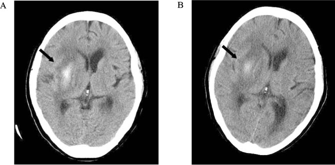
Fig. 1 Brain computed tomography showed intracranial hemorrhage in right basal ganglia with effacement of right lateral ventricle (A: on prior hospital visit). No significant interval change was evident (B: on our hospital visit).
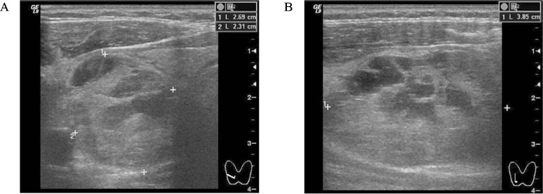
Fig. 2 Neck ultrasonography showed about 2.69 × 2.31 × 3.85 cm sized hypoechoic and septated region in lower pole of right thyroid gland (A. transverse, B. longitudinal).
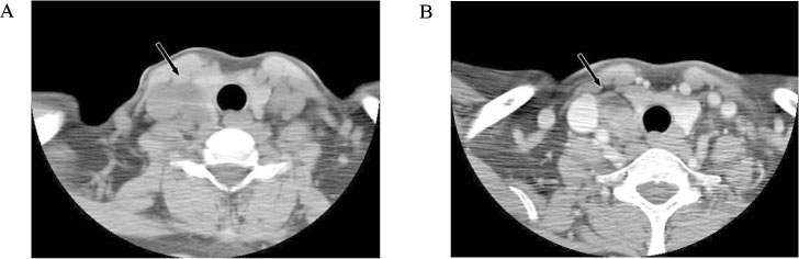
Fig. 3 Neck computed tomography revealed about 3.1 × 1.8 cm sized well defined heterogenous low attenuated mass lateral to right thyroid gland (A. non-enhanced, B. contrast enhanced).
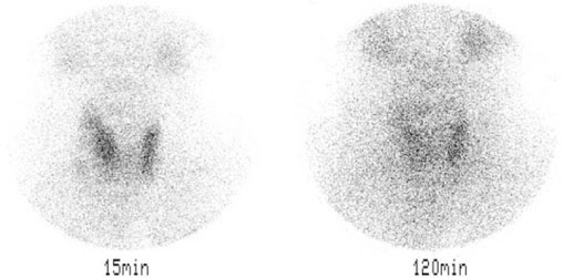
Fig. 4 99mTc-sestamibi scintigraphy showed uneven faint MIBI uptake in lateral and inferior portion of right thyroid lobe after 15 minutes, but no significant MIBI accumulation after 120 minutes.
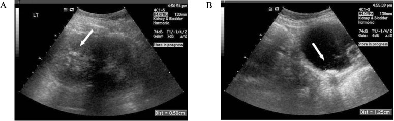
Fig. 5 Abdominal ultrasonography revealed tiny hyperechoic lesion with posterior acoustic shadowing (A). There is about 1.25 cm plate-like hyperechoic lesion in the right posterior wall of urinary bladder (B).
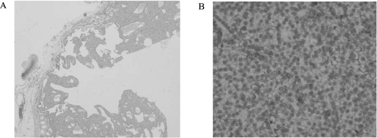
Fig. 6 Microscopic findings showed well-encapsulated mass circumscribed by a rim composed of compressed non-neoplastic parathyroid tissue and relatively thin fibrous connective tissue (A). PTH stain was positive (B). (A. H&E stain ×20, B. Immunohistochemical stain for PTH)
Reference
-
1. Wang CA, Guyton SW. Hyperparathyroid crisis: clinical and pathologic studies of 14 patients. Ann Surg. 1979. 190:782–790.2. Greence EI, Greence JM, Busch RC. Unusual manifestations after removal of parathyroid cyst. J Am Med Assoc. 1952. 150:853–855.3. Gurbuz AT, Peetz ME. Giant mediastinal parathyroid cyst: an unusual cause of hypercalcemic crisis--case report and review of the literature. Surgery. 1996. 120:795–800.4. Chon S, Kim YH, Park JY, Ko KP, Park CY, Kim DY, Woo JT, Kim SW, Kim JW, Kim YS, Go SH. A case of cystic parathyroid adenoma presenting as severe bony lesion. J Kor Soc Endocrinol. 2003. 18:214–220.5. Goris D. Extirpation de trios lobules parathyroidiens kystiques. J Chir Ann Pathol Soc Belge. 1905. 5:394–400.6. Rosenberg J, Orlando R 3rd, Ludwig M, Pyrtek LJ. Parathyroid cysts. Am J Surg. 1982. 143:473–480.7. Layfield LJ. Fine needle aspiration cytology of cystic parathyroid lesions. A cytomorphologic overlap with cystic lesions of the thyroid. Acta Cytol. 1991. 35:447–450.8. Troster M, chiu HF, McLarty TD. Parathyroid cysts: report of a case with ultrastructural observations. Surgery. 1978. 83:238–242.9. Selye H, Ortega MR, Tuchweber B. Experimental production of parathyroid cysts. Am J Pathol. 1964. 45:251–259.10. Rogers LA, Fetter BF, Peete WP. Parathyroid cyst and cystic degeneration of parathyroid adenoma. Arch Pathol. 1969. 88:476–479.11. Calandra DB, Shah KH, Prinz RA, Sullivan H, Hofmann C, Oslapas R, Ernst K, Lawrence AM, Paloyan E. Parathyroid cysts: a report of eleven cases including two associated with hyperparathyroid crisis. Surgery. 1983. 94:887–892.12. Chimenes H, Moreaux J, Lefrileux C, Martin E, Bitan A, Klotz HP. Surgical cure; Determination of parathormone in the fluid of the cystic adenoma. Ann Endocrinol (Paris). 1963. 24:642–649.13. Silverman JF, Khazanie PG, Norris HT, Fore WW. Parathyroid hormone (PTH) assay of parathyroid cysts examined by fine-needle aspiration biopsy. Am J Clin Pathol. 1986. 86:776–780.14. Sen P, Flower N, Papesch M, Davis A, Spedding AV. A benign parathyroid cyst presenting with hoarse voice. J Laryngol Otol. 2000. 114:147–148.15. Clark OH. Parathyroid cysts. Am J Surg. 1978. 135:395–402.16. Mitchell BK, Kinder BK, Cornelius E, Stewart AF. Primary hyperparathyroidism: preoperative localization using technetium-sestamibi scanning. J Clin Endocrinol Metab. 1995. 80:7–10.17. Fisken RA, Heath DA, Bold AM. Hypercalcaemia--a hospital survey. Q J Med. 1980. 49:405–418.18. Fitzpatrick LA, Bilezikian JP. Acute primary hyperparathyroidism. Am J Med. 1987. 82:275–282.19. Pollak MR, Yu AS. Brenner BM, editor. Clinical disturbances of calcium, magnesium, and phosphate metabolism. Brenner and Rector's the kidney. 2004. 7th ed. Philadelphia: WB Saunders Co;1041–1076.20. Mahnensmith RL. Hypercalcemia, hypernatremia, and reversible renal insufficiency. Am J Kidney Dis. 1992. 19:604–608.
- Full Text Links
-
- Actions
-
Cited
- CITED
-
- Close
- Share
-
- Similar articles
-
- A Case of Primary Hyperparathyroidism Associated with Hypercalcemic Crisis and Systemic Calcinosis
- Supernumerary Mediastinal Parathyroid Carcinoma Resulting in Recurrent Primary Hyperparathyroidism and Hypercalcemic Crisis: A Case Report
- Parathyroid Adenoma without Hyperparathyroidism Presenting as a Large Neck Mass
- A Case of Parathyroid Apoplexy of Primary Hyperparathyroidism Presenting as Auditory Hallucinations Accompanied with Hypocalcemia
- Two Cases of Hyperparathyroidism Presenting as Acute Pancreatitis
