Computational Modeling with Fluid-Structure Interaction of the Severe M1 Stenosis Before and After Stenting
- Affiliations
-
- 1Department of Radiology and Research Institute of Radiology, University of Ulsan, College of Medicine, Asan Medical Center, Seoul, Korea. dcsuh@amc.seoul.kr
- 2School of Mechanical and Automotive Engineering University of Ulsan, Ulsan, Korea.
- 3Department of Mechanical Engineering, Dankook University, Korea.
- KMID: 2052077
- DOI: http://doi.org/10.5469/neuroint.2013.8.1.23
Abstract
- PURPOSE
Image-based computational models with fluid-structure interaction (FSI) can be used to perform plaque mechanical analysis in intracranial artery stenosis. We described a process in FSI study applied to symptomatic severe intracranial (M1) stenosis before and after stenting.
MATERIALS AND METHODS
Reconstructed 3D angiography in STL format was transferred to Magics for smoothing of vessel surface and trimming of branch vessels and to HyperMesh for generating tetra volume mesh from triangular surface-meshed 3D angiogram. Computational analysis of blood flow in the blood vessels was performed using the commercial finite element software ADINA Ver 8.5. The distribution of wall shear stress (WSS), peak velocity and pressure was analyzed before and after intracranial stenting.
RESULTS
The wall shear stress distributions from Computational fluid dynamics (CFD) simulation with rigid wall assumption as well as FSI simulation before and after stenting could be compared. The difference of WSS between rigid wall and compliant wall model both in pre- and post-stent case is only minor except at the stenosis region. These WSS values were greatly reduced after stenting to 15~20 Pa at systole and 3~5 Pa at end-diastole in CFD simulation, which are similar in FSI simulations.
CONCLUSION
Our study revealed that FSI simulation before and after intracranial stenting was feasible despite of limited vessel wall dimension and could reveal change of WSS as well as flow velocity and wall pressure.
Keyword
MeSH Terms
Figure
-
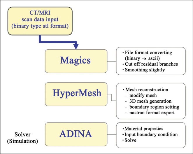
Fig. 1 Analysis process for computational fluid dynamic approach.

Fig. 2 Computational model for FSI in the severe M1 stenosis. Angiograms before (A) and after (B) stenting show relieved severe stenosis of the right M1 and normalization of decreased size and filling of the right MCA branches after stenting. Different material components were assumed for the stented and non-stented vessel in (B).
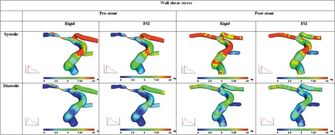
Fig. 3 Wall shear stress distributions before and after stent implantation.
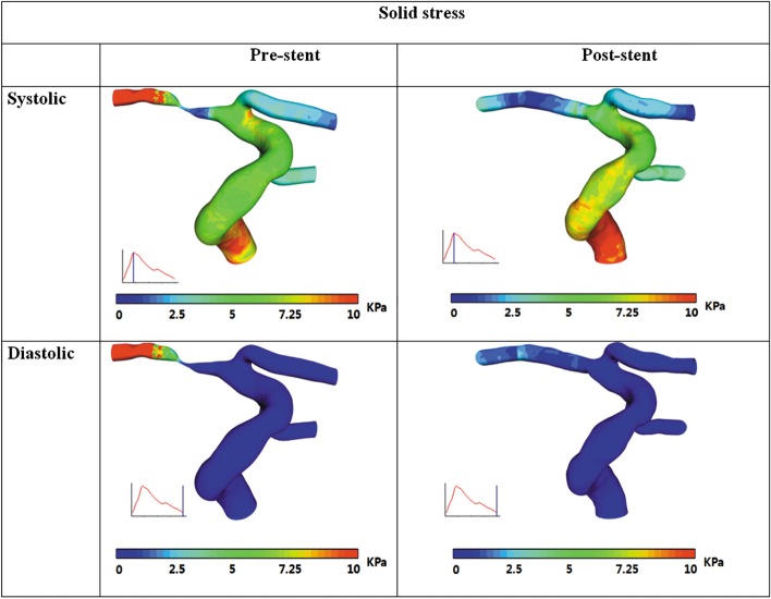
Fig. 4 Solid stress (Von Mises stress) distributions before and after stent implantation.
Cited by 1 articles
-
Computational Fluid Dynamics of Intracranial and Extracranal Arteries using 3-Dimensional Angiography: Technical Considerations with Physician's Point of View
Sung-Tae Park, Kyunghwan Yoon, Young Bae Ko, Dae Chul Suh
Neurointervention. 2013;8(2):92-100. doi: 10.5469/neuroint.2013.8.2.92.
Reference
-
1. Suh DC, Lee SH, Kim KR, Park ST, Lim SM, Kim SJ, et al. Pattern of atherosclerotic carotid stenosis in Korean patients with stroke: different involvement of intracranial versus extracranial vessels. AJNR Am J Neuroradiol. 2003; 24:239–244. PMID: 12591640.2. Gorelick PB, Caplan LR, Hier DB, Parker SL, Patel D. Racial differences in the distribution of anterior circulation occlusive disease. Neurology. 1984; 34:54–59. PMID: 6537853.
Article3. Sacco RL, Kargman DE, Gu Q, Zamanillo MC. Race-ethnicity and determinants of intracranial atherosclerotic cerebral infarction. The Northern Manhattan Stroke Study. Stroke. 1995; 26:14–20. PMID: 7839388.4. Suh DC, Kim JK, Choi JW, Choi BS, Pyun HW, Choi YJ, et al. Intracranial stenting of severe symptomatic intracranial stenosis: results of 100 consecutive patients. AJNR Am J Neuroradiol. 2008; 29:781–785. PMID: 18310234.
Article5. Choi JW, Kim JK, Choi BS, Lim HK, Kim SJ, Kim JS, et al. Angiographic pattern of symptomatic severe M1 stenosis: comparison with presenting symptoms, infarct patterns, perfusion status, and outcome after recanalization. Cerebrovasc Dis. 2010; 29:297–303. PMID: 20090322.
Article6. Suh DC, Sung KB, Cho YS, Choi CG, Lee HK, Lee JH, et al. Transluminal angioplasty for middle cerebral artery stenosis in patients with acute ischemic stroke. AJNR Am J Neuroradiol. 1999; 20:553–558. PMID: 10319958.7. Groen HC, Gijsen FJ, van der Lugt A, Ferguson MS, Hatsukami TS, van der Steen AF, et al. Plaque rupture in the carotid artery is localized at the high shear stress region: a case report. Stroke. 2007; 38:2379–2381. PMID: 17615365.8. Groen HC, Gijsen FJ, van der Lugt A, Ferguson MS, Hatsukami TS, Yuan C, et al. High shear stress influences plaque vulnerability Part of the data presented in this paper were published in Stroke 2007;38:2379-81. Neth Heart J. 2008; 16:280–283. PMID: 18711619.9. Malek AM, Alper SL, Izumo S. Hemodynamic shear stress and its role in atherosclerosis. JAMA. 1999; 282:2035–2042. PMID: 10591386.
Article10. Choi JW, Kim JK, Choi BS, Kim JH, Hwang HJ, Kim JS, et al. Adjuvant revascularization of intracranial artery occlusion with angioplasty and/or stenting. Neuroradiology. 2009; 51:33–43. PMID: 18818910.
Article11. Oh TS, Ko YB, Park ST, Yoon KH, Lee SW, Park JW, et al. Computational Flow Dynamics Study in Severe Carotid Bulb Stenosis with Ulceration. Neurointervention. 2010; 5:97–102.
Article12. Suh DC, Park ST, Oh TS, Park SO, Lim OK, Park SC, et al. High shear stress in systolic phase at the surface of the enhancing plaque is related to symptom presentation of the severe M1 stenosis. Korean J Radiol. 2011; 12:515–518. PMID: 21852914.13. Kim YH, Kim JE, Ito Y, Shih AM, Brott B, Anayiotos A. Hemodynamic analysis of a compliant femoral artery bifurcation model using a fluid structure interaction framework. Ann Biomed Eng. 2008; 36:1753–1763. PMID: 18792781.
Article14. Perktold K, Rappitsch G. Mathematical modeling of arterial blood flow and correlation to atherosclerosis. Technol Health Care. 1995; 3:139–151. PMID: 8749862.
Article15. Gao H, Long Q, Graves M, Gillard JH, Li ZY. Carotid arterial plaque stress analysis using fluid-structure interactive simulation based on in-vivo magnetic resonance images of four patients. J Biomech. 2009; 42:1416–1423. PMID: 19464011.
Article16. Kanyanta V, Ivankovic A, Karac A. Validation of a fluid-structure interaction numerical model for predicting flow transients in arteries. J Biomech. 2009; 42:1705–1712. PMID: 19482285.
Article17. Tang D, Yang C, Mondal S, Liu F, Canton G, Hatsukami TS, et al. A negative correlation between human carotid atherosclerotic plaque progression and plaque wall stress: in vivo MRI-based 2D/3D FSI models. J Biomech. 2008; 41:727–736. PMID: 18191138.
Article18. Leach JR, Rayz VL, Mofrad MR, Saloner D. An efficient two-stage approach for image-based FSI analysis of atherosclerotic arteries. Biomech Model Mechanobiol. 2010; 9:213–223. PMID: 19756798.
Article19. Suh DC, Park ST, Oh TS, Park SO, Lim OK, Park S, et al. High shear stress at the surface of enhancing plaque in the systolic phase is related to the symptom presentation of severe M1 stenosis. Korean J Radiol. 2011; 12:515–518. PMID: 21852914.
Article20. Suh DC, Ko YB, Park ST, Yoon KH, Lim OK, Oh JS, et al. Computational flow dynamics of the severe M1 stenosis before and after stenting. Neurointervention. 2011; 6:13–16. PMID: 22125742.
Article
- Full Text Links
-
- Actions
-
Cited
- CITED
-
- Close
- Share
-
- Similar articles
-
- Computational Flow Dynamics of the Severe M1 Stenosis Before and After Stenting
- Computational Fluid Dynamics Modeling in Aortic Diseases
- High Shear Stress at the Surface of Enhancing Plaque in the Systolic Phase is Related to the Symptom Presentation of Severe M1 Stenosis
- Wingspan Stenting for Symptomatic Severe In-Stent Stenosis of a Closed-Cell Stent after Stent-Assisted Coiling of a Ruptured Intracranial Aneurysm
- Computational modeling of atrial fibrillation
