Imaging Sci Dent.
2015 Sep;45(3):187-192. 10.5624/isd.2015.45.3.187.
Complex dental anomalies in a belatedly diagnosed cleidocranial dysplasia patient
- Affiliations
-
- 1Guanghua School of Stomatology, Guangdong Provincial Key Laboratory of Stomatology, Sun Yat-Sen University, Guangzhou, China. zhaowei3@mail.sysu.edu.cn
- 2Department of Medical Genetics, Zhongshan School of Medicine and Center for Genome Research, Sun Yat-Sen University, Guangzhou, China.
- KMID: 2045027
- DOI: http://doi.org/10.5624/isd.2015.45.3.187
Abstract
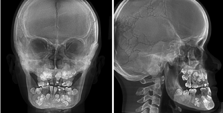
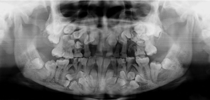
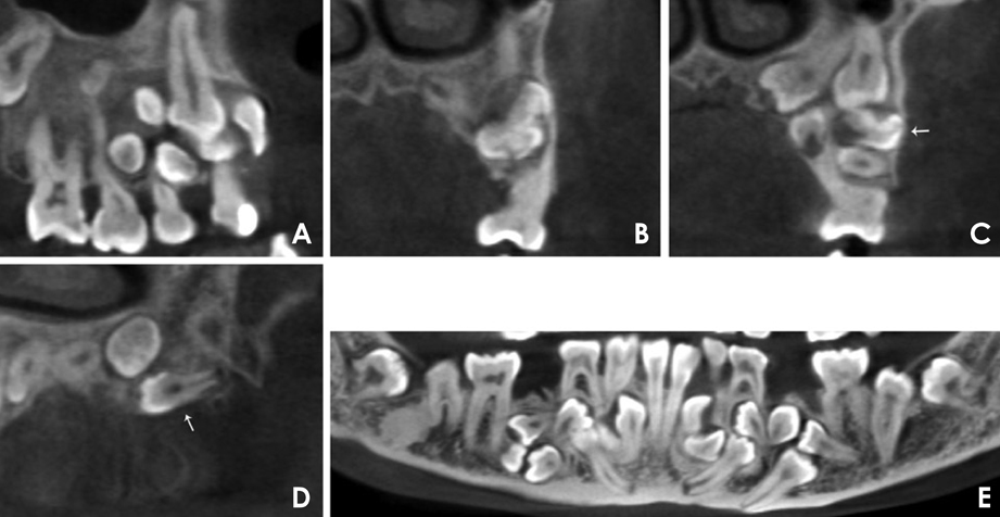
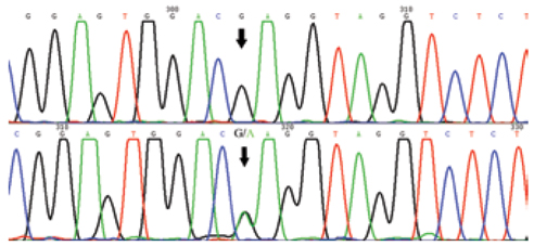
- Cleidocranial dysplasia (CCD) is a rare congenital disorder, typically characterized by persistently open skull sutures, aplastic or hypoplastic clavicles, and supernumerary teeth. Mutations in the gene encoding the runt-related transcription factor 2 (RUNX2) protein are responsible for approximately two thirds of CCD patients. We report a 20-year-old CCD patient presenting not only with typical skeletal changes, but also complex dental anomalies. A previously undiagnosed odontoma, 14 supernumerary teeth, a cystic lesion, and previously unreported fused primary teeth were discovered on cone-beam computed tomography (CBCT) scans. Mutation analysis identified the causal c.578G>A (p.R193Q) mutation in the RUNX2 gene. At 20 years of age, the patient had already missed the optimal period for dental intervention. This report describes the complex dental anomalies in a belatedly diagnosed CCD patient, and emphasizes the significance of CBCT assessment for the detection of dental anomalies and the importance of early treatment to achieve good outcomes.
MeSH Terms
Figure
Cited by 1 articles
-
Case series of cleidocranial dysplasia: Radiographic follow-up study of delayed eruption of impacted permanent teeth
Han-Gyeol Yeom, Won-Jong Park, Eun Joo Choi, Kyung-Hwa Kang, Byung-Do Lee
Imaging Sci Dent. 2019;49(4):307-315. doi: 10.5624/isd.2019.49.4.307.
Reference
-
1. Mundlos S. Cleidocranial dysplasia, clinical and molecular genetics. J Med Genet. 1999; 36:177–182.2. Ott CE, Leschik G, Trotier F, Brueton L, Brunner HG, Brussel W, et al. Deletions of the RUNX2 gene are present in about 10% of individuals with cleidocranial dysplasia. Hum Mutat. 2010; 31:E1587–E1593.
Article3. Karsenty G. Update on the transcriptional control of osteoblast differentiation. Bonekey Osteovision. 2007; 4:164–170.
Article4. Camilleri S, McDonald F. Runx2 and dental development. Eur J Oral Sci. 2006; 114:361–373.
Article5. McNamara CM, O'Riordan BC, Blake M, Sandy JR. Cleidocranial dysplasia: radiological appearances on dental panoramic radiography. Dentomaxillofac Radiol. 1999; 28:89–97.
Article6. Suda N, Hamada T, Hattori M, Torii C, Kosaki K, Moriyama K. Diversity of supernumerary tooth formation in siblings with cleidocranial dysplasia having identical mutation in RUNX2: possible involvement of non-genetic or epigenetic regulation. Orthod Craniofac Res. 2007; 10:222–225.7. Kolokitha OE, Ioannidou I. A 13-year-old Caucasian boy with cleidocranial dysplasia: a case report. BMC Res Notes. 2013; 6:6.
Article8. Wang XP, Fan J. Molecular genetics of supernumerary tooth formation. Genesis. 2011; 49:261–277.
Article9. Chang JY, Wang JT, Wang YP, Liu BY, Sun A, Chiang CP. Odontoma: a clinicopathologic study of 81 cases. J Formos Med Assoc. 2003; 102:876–882.10. Rajab LD, Hamdan MA. Supernumerary teeth: review of the literature and a survey of 152 cases. Int J Paediatr Dent. 2002; 12:244–254.
Article11. Saraçoğlu U, Kurt B, Günhan O, Güven O. MIB-1 expression in odontogenic epithelial rests, epithelium of healthy oral mucosa and epithelium of selected odontogenic cysts. An immunohistochemical study. Int J Oral Maxillofac Surg. 2005; 34:432–435.12. Adaki SR, Yashodadevi BK, Sujatha S, Santana N, Rakesh N, Adaki R. Incidence of cystic changes in impacted lower third molar. Indian J Dent Res. 2013; 24:183–187.
Article13. Muthukumar RS, Arunkumar S, Sadasiva K. Bilateral fusion of mandibular second premolar and supernumerary tooth: a rare case report. J Oral Maxillofac Pathol. 2012; 16:128–130.
Article14. Tsujino K, Yonezu T, Shintani S. Effects of different combinations of fused primary teeth on eruption of the permanent successors. Pediatr Dent. 2013; 35:E64–E67.15. Callea M, Bellacchio E, Di Stazio M, Fattori F, Bertini E, Yavuz I, et al. A case of cleidocranial dysplasia with peculiar dental features: pathogenetic role of the RUNX2 mutation and long term follow-up. Oral Health Dent Manag. 2014; 13:548–551.16. Park TK, Vargervik K, Oberoi S. Orthodontic and surgical management of cleidocranial dysplasia. Korean J Orthod. 2013; 43:248–260.
Article
- Full Text Links
- Actions
-
Cited
- CITED
-
- Close
- Share
- Similar articles
-
- Cranioplasty Using a Modified Split Calvarial Graft Technique in Cleidocranial Dysplasia
- Manifestation and treatment in a cleidocranial dysplasia patient with a RUNX2 (T420I) mutation
- Cleidocranial Dysostosis: One Case Report
- Cleidocranial dysplasia: a preliminary report
- Cleidocranial Dysplasia: Report of a Case

