Imaging Sci Dent.
2015 Sep;45(3):147-151. 10.5624/isd.2015.45.3.147.
The effect of metal artifacts on the identification of vertical root fractures using different fields of view in cone-beam computed tomography
- Affiliations
-
- 1Oral and Maxillofacial Radiology Department, Dental School, Babol University of Medical Science, Babol, Iran. zeinab_nabavi@yahoo.com
- 2Endodontics Department, Dental School, Babol University of Medical Science, Babol, Iran.
- 3Social Department of Health Research Center, Babol University of Medical Science, Babol, Iran.
- KMID: 2045021
- DOI: http://doi.org/10.5624/isd.2015.45.3.147
Abstract
- PURPOSE
The aim of this study was to investigate the effects of metal artifacts on the accurate diagnosis of root fractures using cone-beam computed tomography (CBCT) images with large and small/limited fields of view (FOVs).
MATERIALS AND METHODS
Forty extracted molar and premolar teeth were collected. Access canals were made in all teeth using a rotary system. In half of the teeth, fractures were created by the application of mild pressure with a hammer. The teeth were then randomly put into a wax rim on an acryl base designed in the shape of a mandible. CBCT scans were obtained using a Newtom 5G system with FOVs of 18 cmx16 cm and 6 cmx6 cm. A metal pin was then placed into each tooth, and CBCT imaging was again performed using the same fields of view. All scans were evaluated by two oral and maxillofacial radiologists. The specificity, sensitivity, positive predictive value, negative predictive value, and likelihood ratios (positive and negative) were calculated.
RESULTS
The maximum levels of sensitivity and specificity (100% and 100%, respectively) were observed in smallvolume CBCT scans of teeth without pins. The highest negative predictive value was found in the small-volume group without pins, whereas the positive predictive value was 100% in all groups except the large-volume group with pins.
CONCLUSION
The specificity of CBCT decreased with the presence of a pin in the large-volume group, but not in the small-volume group.
MeSH Terms
Figure
-
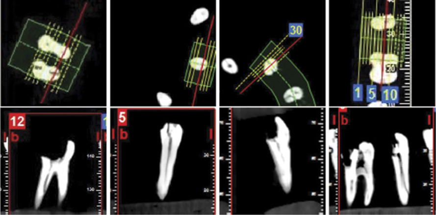
Fig. 1 Fracture lines in axial and cross-sectional cone-beam computed tomography images. A. Large volume without a pin. B. Small volume without a pin. C. Large volume with a pin. D. Small volume with a pin.

Fig. 2 Axial (A) and cross-sectional (B) small-volume cone-beam computed tomography images showing fractures in teeth with a pin. The fracture and the metal atrifact are shown with a straight arrow and a curved arrow, respectively.
Reference
-
1. Katsumata A, Hirukawa A, Okumura S, Naitoh M, Fujishita M, Ariji E, et al. Relationship between density variability and imaging volume size in cone-beam computerized tomographic scanning of the maxillofacial region: an in vitro study. Oral Surg Oral Med Oral Pathol Oral Radiol Endod. 2009; 107:420–425.
Article2. Palomo JM, Kau C, Palomo LB, Hans MG. Three-dimensional cone beam computerized tomography in dentistry. Dent Today. 2006; 25:130–135.3. Bechara BB, Moore WS, McMahan CA, Noujeim M. Metal artefact reduction with cone beam CT: an in vitro study. Dentomaxillofac Radiol. 2012; 41:248–253.4. Ferreira RI, Bahrami G, Isidor F, Wenzel A, Haiter-Neto F, Groppo FC. Detection of vertical root fractures by cone-beam computerized tomography in endodontically treated teeth with fiber-resin and titanium posts: an in vitro study. Oral Surg Oral Med Oral Pathol Oral Radiol. 2013; 115:e49–e57.
Article5. Haghanifar S, Moudi E, Mesgarani A, Bijani A, Abbaszadeh N. A comparative study of cone-beam computed tomography and digital periapical radiography in detecting mandibular molars root perforations. Imaging Sci Dent. 2014; 44:115–119.
Article6. Hassan B, Metska ME, Ozok AR, van der Stelt P, Wesselink PR. Comparison of five cone beam computed tomography systems for the detection of vertical root fractures. J Endod. 2010; 36:126–129.
Article7. Bornstein MM, Wölner-Hanssen AB, Sendi P, von Arx T. Comparison of intraoral radiography and limited cone beam computed tomography for the assessment of root-fractured permanent teeth. Dent Traumatol. 2009; 25:571–577.
Article8. Costa FF, Gaia BF, Umetsubo OS, Pinheiro LR, Tortamano IP, Cavalcanti MG. Use of large-volume cone-beam computed tomography in identification and localization of horizontal root fracture in the presence and absence of intracanal metallic post. J Endod. 2012; 38:856–859.
Article9. Pauwels R, Stamatakis H, Bosmans H, Bogaerts R, Jacobs R, Horner K, et al. Quantification of metal artifacts on cone beam computed tomography images. Clin Oral Implants Res. 2013; 24:94–99.
Article10. Scarfe WC, Levin MD, Gane D, Farman AG. Use of cone beam computed tomography in endodontics. Int J Dent. 2009; 2009:634567.
Article11. Costa FF, Gaia BF, Umetsubo OS, Cavalcanti MG. Detection of horizontal root fracture with small-volume cone-beam computed tomography in the presence and absence of intracanal metallic post. J Endod. 2011; 37:1456–1459.
Article12. Moudi E, Haghanifar S, Madani Z, Alhavaz A, Bijani A, Bagheri M. Assessment of vertical root fracture using cone-beam computed tomography. Imaging Sci Dent. 2014; 44:37–41.
Article13. Iikubo M, Kobayashi K, Mishima A, Shimoda S, Daimaruya T, Igarashi C, et al. Accuracy of intraoral radiography, multidetector helical CT, and limited cone-beam CT for the detection of horizontal tooth root fracture. Oral Surg Oral Med Oral Pathol Oral Radiol Endod. 2009; 108:e70–e74.
Article14. Kamburoğlu K, Murat S, Yüksel SP, Cebeci AR, Horasan S. Detection of vertical root fracture using cone-beam computerized tomography: an in vitro assessment. Oral Surg Oral Med Oral Pathol Oral Radiol Endod. 2010; 109:e74–e81.
Article15. Wang P, Yan XB, Lui DG, Zhang WL, Zhang Y, Ma XC. Detection of dental root fractures by using cone-beam computed tomography. Dentomaxillofac Radiol. 2011; 40:290–298.
Article16. Wenzel A, Haiter-Neto F, Frydenberg M, Kirkevang LL. Variable-resolution cone-beam computerized tomography with enhancement filtration compared with intraoral photostimulable phosphor radiography in detection of transverse root fractures in an in vitro model. Oral Surg Oral Med Oral Pathol Oral Radiol Endod. 2009; 108:939–945.
Article
- Full Text Links
-
- Actions
-
Cited
- CITED
-
- Close
- Share
-
- Similar articles
-
- Effect of object position in the field of view and application of a metal artifact reduction algorithm on the detection of vertical root fractures on cone-beam computed tomography scans: An in vitro study
- Effect of cone-beam computed tomography metal artefact reduction on incomplete subtle vertical root fractures
- Assessment of vertical root fracture using cone-beam computed tomography
- Detection of maxillary second molar with two palatal roots using cone beam computed tomography: a case report
- Management of root canal perforation by using cone-beam computed tomography
