Effect of object position in the field of view and application of a metal artifact reduction algorithm on the detection of vertical root fractures on cone-beam computed tomography scans: An in vitro study
- Affiliations
-
- 1Department of Maxillofacial Radiology, Faculty of Dentistry, Guilan University of Medical Sciences, Rasht, Iran.
- 2Dental Sciences Research Center, Department of Maxillofacial Radiology, Faculty of Dentistry, Guilan University of Medical Sciences, Rasht, Iran. zahradalili@yahoo.com
- 3Dental Sciences Research Center, Department of Endodontics, Faculty of Dentistry, Guilan University of Medical Sciences, Rasht, Iran.
- KMID: 2450164
- DOI: http://doi.org/10.5624/isd.2018.48.4.245
Abstract
- PURPOSE
To assess the effects of object position in the field of view (FOV) and application of a metal artifact reduction (MAR) algorithm on the diagnostic accuracy of cone-beam computed tomography (CBCT) for the detection of vertical root fractures (VRFs).
MATERIALS AND METHODS
Sixty human single-canal premolars received root canal treatment. VRFs were induced in 30 endodontically treated teeth. The teeth were then divided into 4 groups, with 2 groups receiving metal posts and the remaining 2 only having an empty post space. The roots from different groups were mounted in a phantom made of cow rib bone, and CBCT scans were obtained for the 4 different groups. Three observers evaluated the images independently.
RESULTS
The highest frequency of correct diagnoses of VRFs was obtained with the object positioned centrally in the FOV, using the MAR algorithm. Peripheral positioning of the object without the MAR algorithm yielded the highest sensitivity for the first observer (66.7%). For the second and third observers, a central position improved sensitivity, with or without the MAR algorithm. In the presence of metal posts, central positioning of the object in the FOV significantly increased the diagnostic sensitivity and accuracy compared to peripheral positioning.
CONCLUSION
Diagnostic accuracy was higher with central positioning than with peripheral positioning, irrespective of whether the MAR algorithm was applied. However, the effect of the MAR algorithm was more significant with central positioning than with peripheral positioning of the object in the FOV. The clinical experience and expertise of the observers may serve as a confounder in this respect.
MeSH Terms
Figure
-

Fig. 1 A photograph showing the phantom fabricated for the placement of teeth after tooth insertion.
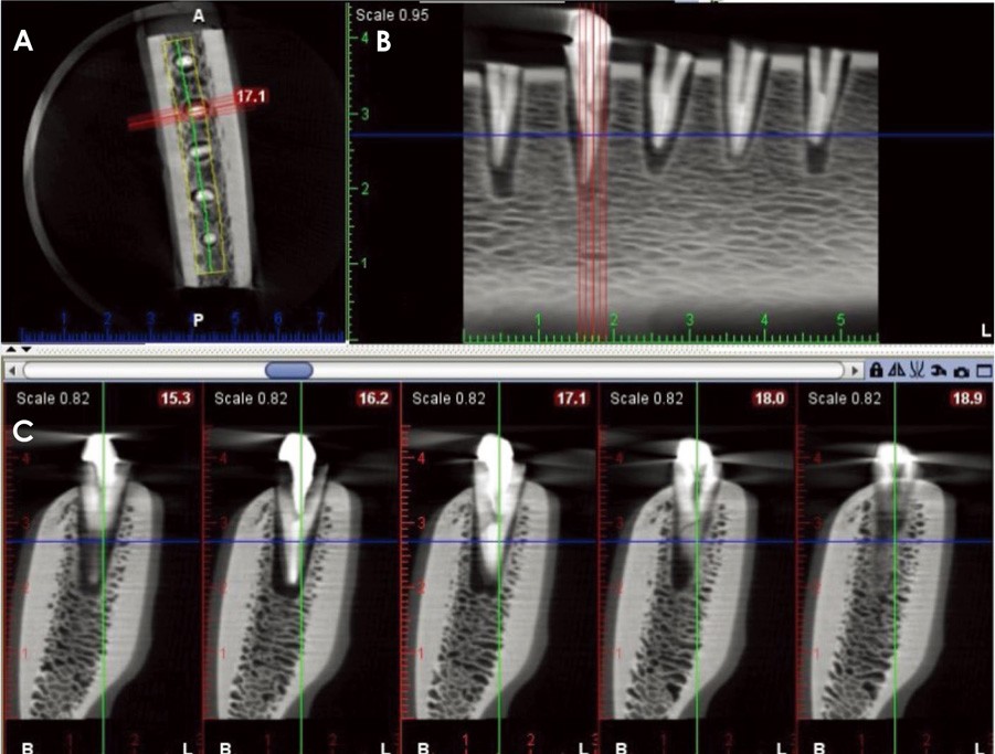
Fig. 2 Cone-beam computed tomographic images with central positioning of the object in the field of view. An axial image (A) and cross-sectional images (B and C) reveal root fracture of the left second tooth.
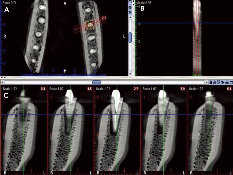
Fig. 3 Cone-beam computed tomographic images with peripheral positioning of the object in the field of view. An axial image (A) and cross-sectional images (B and C) reveal root fracture of the left second tooth.
Cited by 2 articles
-
Magnitude of beam-hardening artifacts produced by gutta-percha and metal posts on cone-beam computed tomography with varying tube current
Hugo Gaêta-Araujo, Eduarda Helena Leandro Nascimento, Rocharles Cavalcante Fontenele, Arthur Xavier Maseti Mancini, Deborah Queiroz Freitas, Christiano Oliveira-Santos
Imaging Sci Dent. 2020;50(1):1-7. doi: 10.5624/isd.2020.50.1.1.Effect of slice inclination and object position within the field of view on the measurement accuracy of potential implant sites on cone-beam computed tomography
Bardia Vadiati Saberi, Negar Khosravifard, Alireza Nourzadeh
Imaging Sci Dent. 2020;50(1):37-43. doi: 10.5624/isd.2020.50.1.37.
Reference
-
1. Kamburoğlu K, Murat S, Yüksel SP, Cebeci AR, Horasan S. Detection of vertical root fracture using cone-beam computerized tomography: an in vitro assessment. Oral Surg Oral Med Oral Pathol Oral Radiol Endod. 2010; 109:e74–e81.
Article2. Tamse A. Vertical root fractures in endodontically treated teeth: diagnostic signs and clinical management. Endod Topics. 2006; 13:84–94.
Article3. Khedmat S, Rouhi N, Drage N, Shokouhinejad N, Nekoofar MH. Evaluation of three imaging techniques for the detection of vertical root fractures in the absence and presence of gutta-percha root fillings. Int Endod J. 2012; 45:1004–1009.
Article4. Patel S, Dawood A, Ford TP, Whaites E. The potential applications of cone beam computed tomography in the management of endodontic problems. Int Endod J. 2007; 40:818–830.
Article5. Pauwels R, Stamatakis H, Bosmans H, Bogaerts R, Jacobs R, Horner K, et al. Quantification of metal artifacts on cone beam computed tomography images. Clin Oral Implants Res. 2013; 24:Suppl A100. 94–99.
Article6. Bernardes RA, de Moraes IG, Húngaro Duarte MA, Azevedo BC, de Azevedo JR, Bramante CM. Use of cone-beam volumetric tomography in the diagnosis of root fractures. Oral Surg Oral Med Oral Pathol Oral Radiol Endod. 2009; 108:270–277.
Article7. Ferreira RI, Bahrami G, Isidor F, Wenzel A, Haiter-Neto F, Groppo FC. Detection of vertical root fractures by cone-beam computerized tomography in endodontically treated teeth with fiber-resin and titanium posts: an in vitro study. Oral Surg Oral Med Oral Pathol Oral Radiol. 2013; 115:e49–e57.
Article8. Youssefzadeh S, Gahleitner A, Dorffner R, Bernhart T, Kainberger FM. Dental vertical root fractures: value of CT in detection. Radiology. 1999; 210:545–549.
Article9. Moudi E, Haghanifar S, Madani Z, Alhavaz A, Bijani A, Bagheri M. Assessment of vertical root fracture using cone-beam computed tomography. Imaging Sci Dent. 2014; 44:37–41.
Article10. Kajan ZD, Taromsari M. Value of cone beam CT in detection of dental root fractures. Dentomaxillofac Radiol. 2012; 41:3–10.
Article11. Wang P, Yan XB, Lui DG, Zhang WL, Zhang Y, Ma XC. Detection of dental root fractures by using cone-beam computed tomography. Dentomaxillofac Radiol. 2011; 40:290–298.
Article12. Bornstein MM, Wölner-Hanssen AB, Sendi P, von Arx T. Comparison of intraoral radiography and limited cone beam computed tomography for the assessment of root-fractured permanent teeth. Dent Traumatol. 2009; 25:571–577.
Article13. Parsa A, Ibrahim N, Hassan B, Syriopoulos K, van der Stelt P. Assessment of metal artefact reduction around dental titanium implants in cone beam CT. Dentomaxillofac Radiol. 2014; 43:20140019.
Article14. Pauwels R, Jacobs R, Bogaerts R, Bosmans H, Panmekiate S. Reduction of scatter-induced image noise in cone beam computed tomography: effect of field of view size and position. Oral Surg Oral Med Oral Pathol Oral Radiol. 2016; 121:188–195.
Article15. Queiroz PM, Santaella GM, da Paz TD, Freitas DQ. Evaluation of a metal artefact reduction tool on different positions of a metal object in the FOV. Dentomaxillofac Radiol. 2017; 46:20160366.
Article16. Bechara BB, Moore WS, McMahan CA, Noujeim M. Metal artefact reduction with cone beam CT: an in vitro study. Dentomaxillofac Radiol. 2012; 41:248–253.17. Bezerra IS, Neves FS, Vasconcelos TV, Ambrosano GM, Freitas DQ. Influence of the artefact reduction algorithm of Picasso Trio CBCT system on the diagnosis of vertical root fractures in teeth with metal posts. Dentomaxillofac Radiol. 2015; 44:20140428.
Article18. Barrett JF, Keat N. Artifacts in CT: recognition and avoidance. Radiographics. 2004; 24:1679–1691.
Article19. Özer SY. Detection of vertical root fractures of different thicknesses in endodontically enlarged teeth by cone beam computed tomography versus digital radiography. J Endod. 2010; 36:1245–1249.20. Junqueira RB, Verner FS, Campos CN, Devito KL, do Carmo AM. Detection of vertical root fractures in the presence of intracanal metallic post: a comparison between periapical radiography and cone-beam computed tomography. J Endod. 2013; 39:1620–1624.
Article21. Melo SL, Haiter-Neto F, Correa LR, Scarfe WC, Farman AG. Comparative diagnostic yield of cone beam CT reconstruction using various software programs on the detection of vertical root fractures. Dentomaxillofac Radiol. 2013; 42:20120459.
Article22. Bechara B, Alex McMahan C, Moore WS, Noujeim M, Teixeira FB, Geha H. Cone beam CT scans with and without artefact reduction in root fracture detection of endodontically treated teeth. Dentomaxillofac Radiol. 2013; 42:20120245.
Article23. Dalili Kajan Z, Taramsari M, Khosravi Fard N, Khaksari F, Moghasem Hamidi F. The efficacy of metal artifact reduction mode in cone-beam computed tomography images on diagnostic accuracy of root fractures in teeth with intracanal posts. Iran Endod J. 2018; 13:47–53.24. Iikubo M, Nishioka T, Okura S, Kobayashi K, Sano T, Katsumata A, et al. Influence of voxel size and scan field of view on fracture-like artifacts from gutta-percha obturated endodontically treated teeth on cone-beam computed tomography images. Oral Surg Oral Med Oral Pathol Oral Radiol. 2016; 122:631–637.
Article25. Queiroz PM, Oliveira ML, Groppo FC, Haiter-Neto F, Freitas DQ. Evaluation of metal artefact reduction in cone-beam computed tomography images of different dental materials. Clin Oral Investig. 2018; 22:419–423.
Article26. Salineiro FCS, Kobayashi-Velasco S, Braga MM, Cavalcanti MGP. Radiographic diagnosis of root fractures: a systematic review, meta-analyses and sources of heterogeneity. Dentomaxillofac Radiol. 2017; 46:20170400.
Article27. Katsumata A, Hirukawa A, Okumura S, Naitoh M, Fujishita M, Ariji E, et al. Relationship between density variability and imaging volume size in cone-beam computerized tomographic scanning of the maxillofacial region: an in vitro study. Oral Surg Oral Med Oral Pathol Oral Radiol Endod. 2009; 107:420–425.
Article28. van Daatselaar AN, Dunn SM, Spoelder HJ, Germans DM, Renambot L, Bal HE, et al. Feasibility of local CT of dental tissues. Dentomaxillofac Radiol. 2003; 32:173–180.
Article29. Katsumata A, Hirukawa A, Okumura S, Naitoh M, Fujishita M, Ariji E, et al. Effects of image artifacts on gray-value density in limited-volume cone-beam computerized tomography. Oral Surg Oral Med Oral Pathol Oral Radiol Endod. 2007; 104:829–836.
Article
- Full Text Links
-
- Actions
-
Cited
- CITED
-
- Close
- Share
-
- Similar articles
-
- Effect of a metal artifact reduction algorithm on cone-beam computed tomography scans of titanium and zirconia implants within and outside the field of view
- Effect of cone-beam computed tomography metal artefact reduction on incomplete subtle vertical root fractures
- The effect of metal artifacts on the identification of vertical root fractures using different fields of view in cone-beam computed tomography
- Metal artifact production and reduction in CBCT with different numbers of basis images
- Vertical root fracture diagnosis in teeth with metallic posts: Impact of metal artifact reduction and sharpening filters
