Imaging Sci Dent.
2018 Mar;48(1):41-44. 10.5624/isd.2018.48.1.41.
Metal artifact production and reduction in CBCT with different numbers of basis images
- Affiliations
-
- 1Department of Oral Diagnosis, Area of Oral Radiology, Piracicaba Dental School, University of Campinas, Piracicaba, Sao Paulo, Brazil. gustavoms@live.com
- 2Department of Physiological Sciences, Area of Pharmacology, Piracicaba Dental School, University of Campinas, Piracicaba, Sao Paulo, Brazil.
- KMID: 2406981
- DOI: http://doi.org/10.5624/isd.2018.48.1.41
Abstract
- PURPOSE
To evaluate the effect of different numbers of basis images and the use of metal artifact reduction (MAR) on the production and reduction of artifacts in cone-beam computed tomography images.
MATERIALS AND METHODS
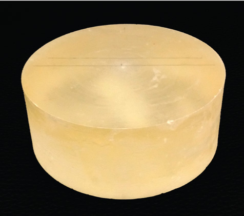
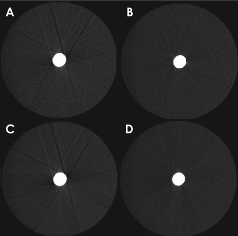
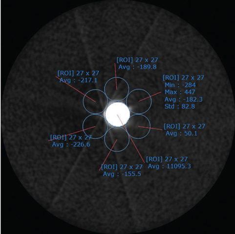
An acrylic resin phantom with a metal alloy sample was scanned, with 450 or 720 basis images and with or without MAR. Standard deviation values for the test areas (around the metal object) were obtained as a way of measuring artifact production. Two-way analysis of variance was used with a 5% significance level.
RESULTS
There was no significant difference in artifact production among the images obtained with different numbers of basis images without MAR (P=.985). MAR significantly reduced artifact production in the test areas only in the protocol using 720 basis images (P=.017). The protocol using 450 basis images with MAR showed no significant difference in artifact production when compared to the protocol using 720 basis images with MAR (P=.579).
CONCLUSION
Protocols with a smaller number of basis images and with MAR activated are preferable for minimizing artifact production in tomographic images without exposing the patient to a greater radiation dose.
Figure
Reference
-
1. Scarfe WC, Farman AG, Sukovic P. Clinical applications of cone-beam computed tomography in dental practice. J Can Dent Assoc. 2006; 72:75–80.2. Vande Berg B, Malghem J, Maldague B, Lecouvet F. Multi-detector CT imaging in the postoperative orthopedic patient with metal hardware. Eur J Radiol. 2006; 60:470–479.
Article3. Draenert FG, Coppenrath E, Herzog P, Müller S, Mueller-Lisse UG. Beam hardening artefacts occur in dental implant scans with the NewTom cone beam CT but not with the dental 4-row multidetector CT. Dentomaxillofac Radiol. 2007; 36:198–203.4. Schulze R, Heil U, Gross D, Bruellmann DD, Dranischnikow E, Schwanecke U, et al. Artefacts in CBCT: a review. Dentomaxillofac Radiol. 2011; 40:265–273.
Article5. Kamburoğlu K, Kolsuz E, Murat S, Eren H, Yüksel S, Paksoy CS. Assessment of buccal marginal alveolar peri-implant and periodontal defects using a cone beam CT system with and without the application of metal artefact reduction mode. Dentomaxillofac Radiol. 2013; 42:20130176.
Article6. Bechara B, McMahan CA, Nasseh I, Geha H, Hayek E, Khawam G, et al. Number of basis images effect on detection of root fractures in endodontically treated teeth using a cone beam computed tomography machine: an in vitro study. Oral Surg Oral Med Oral Pathol Oral Radiol. 2013; 115:676–681.
Article7. Bezerra IS, Neves FS, Vasconcelos TV, Ambrosano GM, Freitas DQ. Influence of the artefact reduction algorithm of Picasso Trio CBCT system on the diagnosis of vertical root fractures in teeth with metal posts. Dentomaxillofac Radiol. 2015; 44:20140428.
Article8. Scarfe WC, Farman AG. What is cone-beam CT and how does it work? Dent Clin North Am. 2008; 52:707–730.
Article9. Wenzel A, Sewerin I. Sources of noise in digital subtraction radiography. Oral Surg Oral Med Oral Pathol. 1991; 71:503–508.
Article10. Spin-Neto R, Matzen LH, Schropp L, Gotfredsen E, Wenzel A. Movement characteristics in young patients and the impact on CBCT image quality. Dentomaxillofac Radiol. 2016; 45:20150426.
Article
- Full Text Links
- Actions
-
Cited
- CITED
-
- Close
- Share
- Similar articles
-
- Influence of CBCT metal artifact reduction on vertical radicular fracture detection
- Theoretical Investigation of Metal Artifact Reduction Based on Sinogram Normalization in Computed Tomography
- Reduction of Metal Artifact around Titanium Alloy-based Pedicle Screws on CT Scan Images: An Approach using a Digital Image Enhancement Technique
- Value and Clinical Application of Orthopedic Metal Artifact Reduction Algorithm in CT Scans after Orthopedic Metal Implantation
- Does the metal artifact reduction algorithm activation mode influence the magnitude of artifacts in CBCT images?


