Preoperative Weakness and Demyelination of the Corticospinal Tract in Meningioma Patients : Changes in Diffusion Parameters Using Diffusion Tensor Imaging
- Affiliations
-
- 1Department of Neurosurgery, Seoul Paik Hospital, Inje University College of Medicine, Seoul, Korea.
- 2Department of Neurosurgery, Seoul National University College of Medicine, Seoul, Korea. chungc@snu.ac.kr
- 3Neuroscience Research Institute, Seoul National University Medical Research Center, Seoul, Korea.
- 4Clinical Research Institute, Seoul National University Hospital, Seoul, Korea.
- KMID: 2018032
- DOI: http://doi.org/10.3340/jkns.2014.55.5.267
Abstract
OBJECTIVE
Differentiation of demyelination in white matter from axonal damage can be determined using diffusion tensor imaging (DTI). In this study using meningioma patients an attempt was made to evaluate the relationship between preoperative weakness and the changes of diffusion parameters in the corticospinal tract (CST) using DTI.
METHODS
Twenty-six patients with meningioma were enrolled in this study. Eleven of them suffered from objective motor weakness and were classified as Group 1. The remaining 15 patients did not present motor weakness and were classified as Group 2. Fiber tractography and CST diffusion parameters were obtained using DTIStudio. The ratios (lesion side mean value/contralateral side mean value) of CST diffusion parameters were compared with 1.0 as a test value using a one-sample t-test.
RESULTS
In Group 1, fractional anisotropy (FA), tensor trace (TT), and radial diffusivity (RD, lambda2 and lambda3) of the CST were significantly different between two hemispheres, but axial diffusivity (AD, lambda1) of the CST was not significantly different between two hemispheres. In Group 2, FA and lambda3 of CST did not differ significantly between the hemispheres. In Group 2, TT, lambda1, and lambda2 of CST in the ipsilateral hemisphere were significantly higher than those of the unaffected hemisphere. However, the differences were small.
CONCLUSION
Motor weakness was related to a low FA and high TT resulting from increased RD of the CST fibers. CST diffusion changes in patients with weakness are similar to those for demyelination.
MeSH Terms
Figure
-
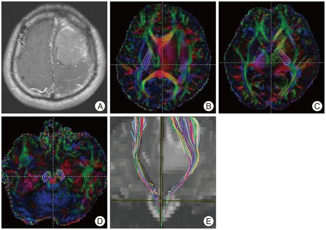
Fig. 1 Corticospinal tract (CST) fiber tracking in a 37-year-old man with benign meningioma. A 37-year-old man presented with seizure and right-upper-extremity weakness. A T1-enhanced image shows a slightly enhanced mass in the left frontal lobe (A). The posterior limb of the internal capsule is selected as the seed region of interest (ROI) (B and C). The ipsilateral cerebral peduncle is selected as the target ROI (D). The tractography image shows the CST on a coronal T2-weighted image (E).
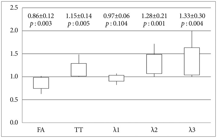
Fig. 2 Ratios (lesion/contralateral) of the diffusion parameters (FA, TT, λ1, λ2, and λ3) for the CST in meningioma patients with weakness (Group 1). y-axis : ratio of diffusion parameters for the CST in the two hemispheres, FA : fractional anisotropy, TT : tensor trace, 1 : primary eigenvalue, λ2 : second eigenvalue, λ3 : third eigenvalue.

Fig. 3 Ratios (lesion/contralateral) of diffusion parameters (FA, TT, λ1, λ2, and λ3) for the CST in meningioma patients without weakness (Group 2). y-axis : ratio of diffusion parameters for the CST in the two hemispheres, FA : fractional anisotropy, TT : tensor trace, λ1 : primary eigenvalue, λ2 : second eigenvalue, λ3 : third eigenvalue.
Reference
-
1. Bello L, Gambini A, Castellano A, Carrabba G, Acerbi F, Fava E, et al. Motor and language DTI Fiber Tracking combined with intraoperative subcortical mapping for surgical removal of gliomas. Neuroimage. 2008; 39:369–382. PMID: 17911032.
Article2. Berman JI, Mukherjee P, Partridge SC, Miller SP, Ferriero DM, Barkovich AJ, et al. Quantitative diffusion tensor MRI fiber tractography of sensorimotor white matter development in premature infants. Neuroimage. 2005; 27:862–871. PMID: 15978841.
Article3. Budde MD, Xie M, Cross AH, Song SK. Axial diffusivity is the primary correlate of axonal injury in the experimental autoimmune encephalomyelitis spinal cord : a quantitative pixelwise analysis. J Neurosci. 2009; 29:2805–2813. PMID: 19261876.
Article4. Field AS, Alexander AL, Wu YC, Hasan KM, Witwer B, Badie B. Diffusion tensor eigenvector directional color imaging patterns in the evaluation of cerebral white matter tracts altered by tumor. J Magn Reson Imaging. 2004; 20:555–562. PMID: 15390227.
Article5. Helton KJ, Phillips NS, Khan RB, Boop FA, Sanford RA, Zou P, et al. Diffusion tensor imaging of tract involvement in children with pontine tumors. AJNR Am J Neuroradiol. 2006; 27:786–793. PMID: 16611765.6. Kim CH, Chung CK, Kim JS, Jahng TA, Lee JH, Song IC. Use of diffusion tensor imaging to evaluate weakness. J Neurosurg. 2007; 106:111–118. PMID: 17236496.
Article7. Loy DN, Kim JH, Xie M, Schmidt RE, Trinkaus K, Song SK. Diffusion tensor imaging predicts hyperacute spinal cord injury severity. J Neurotrauma. 2007; 24:979–990. PMID: 17600514.
Article8. Mori S, Frederiksen K, van Zijl PC, Stieltjes B, Kraut MA, Solaiyappan M, et al. Brain white matter anatomy of tumor patients evaluated with diffusion tensor imaging. Ann Neurol. 2002; 51:377–380. PMID: 11891834.
Article9. Nagesh V, Tsien CI, Chenevert TL, Ross BD, Lawrence TS, Junick L, et al. Radiation-induced changes in normal-appearing white matter in patients with cerebral tumors : a diffusion tensor imaging study. Int J Radiat Oncol Biol Phys. 2008; 70:1002–1010. PMID: 18313524.
Article10. Nilsson D, Rutka JT, Snead OC 3rd, Raybaud CR, Widjaja E. Preserved structural integrity of white matter adjacent to low-grade tumors. Childs Nerv Syst. 2008; 24:313–320. PMID: 17960393.
Article11. Okada T, Mikuni N, Miki Y, Kikuta K, Urayama S, Hanakawa T, et al. Corticospinal tract localization : integration of diffusion-tensor tractography at 3-T MR imaging with intraoperative white matter stimulation mapping-preliminary results. Radiology. 2006; 240:849–857. PMID: 16857980.
Article12. Reich DS, Smith SA, Jones CK, Zackowski KM, van Zijl PC, Calabresi PA, et al. Quantitative characterization of the corticospinal tract at 3T. AJNR Am J Neuroradiol. 2006; 27:2168–2178. PMID: 17110689.13. Salat DH, Tuch DS, Greve DN, van der Kouwe AJ, Hevelone ND, Zaleta AK, et al. Age-related alterations in white matter microstructure measured by diffusion tensor imaging. Neurobiol Aging. 2005; 26:1215–1227. PMID: 15917106.
Article14. Schonberg T, Pianka P, Hendler T, Pasternak O, Assaf Y. Characterization of displaced white matter by brain tumors using combined DTI and fMRI. Neuroimage. 2006; 30:1100–1111. PMID: 16427322.
Article15. Smits M, Vernooij MW, Wielopolski PA, Vincent AJ, Houston GC, van der Lugt A. Incorporating functional MR imaging into diffusion tensortractography in the preoperative assessment of the corticospinal tract in patients with brain tumors. AJNR Am J Neuroradiol. 2007; 28:1354–1361. PMID: 17698540.
Article16. Song SK, Sun SW, Ju WK, Lin SJ, Cross AH, Neufeld AH. Diffusion tensor imaging detects and differentiates axon and myelin degeneration in mouse optic nerve after retinal ischemia. Neuroimage. 2003; 20:1714–1722. PMID: 14642481.
Article17. Song SK, Sun SW, Ramsbottom MJ, Chang C, Russell J, Cross AH. Dysmyelination revealed through MRI as increased radial (but unchanged axial) diffusion of water. Neuroimage. 2002; 17:1429–1436. PMID: 12414282.
Article18. Stadlbauer A, Ganslandt O, Buslei R, Hammen T, Gruber S, Moser E, et al. Gliomas : histopathologic evaluation of changes in directionality and magnitude of water diffusion at diffusion-tensor MR imaging. Radiology. 2006; 240:803–810. PMID: 16926329.
Article
- Full Text Links
-
- Actions
-
Cited
- CITED
-
- Close
- Share
-
- Similar articles
-
- Mini-Review of Studies Reporting the Repeatability and Reproducibility of Diffusion Tensor Imaging
- Principle and Experiments in Diffusion Tensor Imaging
- Disruption of the Corticoreticular Tract in Pediatric Patients With Trunk Instability: A Diffusion Tensor Tractography Study
- Optimal Factors of Diffusion Tensor Imaging Predicting Corticospinal Tract Injury in Patients with Brain Tumors
- Clinical Usefulness of Diffusion Tensor Image Tractography in Stroke Patients: Report of two cases
