Ann Rehabil Med.
2017 Dec;41(6):1093-1099. 10.5535/arm.2017.41.6.1093.
Disruption of the Corticoreticular Tract in Pediatric Patients With Trunk Instability: A Diffusion Tensor Tractography Study
- Affiliations
-
- 1Department of Physical Medicine and Rehabilitation, Yeungnam University College of Medicine, Daegu, Korea. nocturne27@naver.com
- KMID: 2400296
- DOI: http://doi.org/10.5535/arm.2017.41.6.1093
Abstract
- The authors report the diffusion tensor tractography (DTT) findings of three pediatric patients with gait dysfunction and corticoreticular tract (CRT) disruption. All three patients showed unilateral trunk instability, but they did not show any spasticity or weakness of the distal extremities. Clinical evaluation of trunk instability using a Trunk Control Measurement Scale (TCMS) revealed that the more affected side had a lower score than the contralateral side. DTT showed disrupted CRTs in hemispheres contralateral to the hemiparetic sides, which were associated with unilateral proximal instability, although conventional MRI showed no abnormal lesion explaining the hemiplegic symptom. Compared to the results in age-matched controls, these three patients had decreased values of fractional anisotropy (FA) and tract volumes (TV) of the affected CRTs, and these values were also decreased compared to those in the contralateral side. On the other hand, values of FA and TV of the corticospinal tracts on the ipsilateral and contralateral sides were only marginally different. In conclusion, diffusion tensor imaging can be helpful for investigating the state of the CRT in pediatric patients with trunk instability and gait dysfunction.
Keyword
MeSH Terms
Figure
-
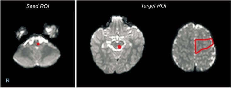
Fig. 1 Regions of interest (ROIs) were set for the corticoreticular tract (seed ROI, reticular formation in the medulla; first target ROI, midbrain tegmentum; second target ROI, Brodmann area 6).
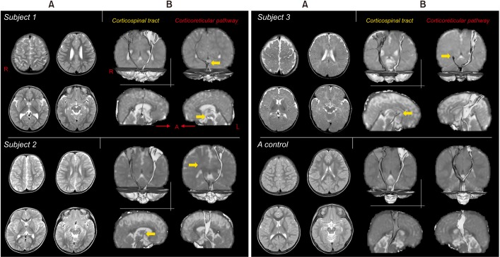
Fig. 2 (A) T2-weighted axial magnetic resonance images. (B) Diffusion tensor tractography of the corticospinal tract (CST) and the corticoreticular tract (CRT). Bilateral CSTs were preserved in all three patients. Subject 1 showed disruption of the left CRT, whereas subjects 2 and 3 showed disruption of the right CRTs. Control subjects showed preserved integrity of the CST and CRT.
Reference
-
1. Lessek AM. The pyramidal tract; basic considerations of corticospinal neurons. Res Publ Assoc Res Nerv Ment Dis. 1948; 27:106–128. PMID: 18106853.2. Matsuyama K, Mori F, Nakajima K, Drew T, Aoki M, Mori S. Locomotor role of the corticoreticular-reticulospinal-spinal interneuronal system. Prog Brain Res. 2004; 143:239–249. PMID: 14653169.
Article3. Freund H-J, Hummelsheim H. Premotor cortex in man: evidence for innervation of proximal limb muscles. Exp Brain Res. 1984; 53:479–482. PMID: 6705877.
Article4. Yeo SS, Chang MC, Kwon YH, Jung YJ, Jang SH. Corticoreticular pathway in the human brain: diffusion tensor tractography study. Neurosci Lett. 2012; 508:9–12. PMID: 22197953.
Article5. Yeo SS, Jang SH. Recovery of an injured corticospinal tract and an injured corticoreticular pathway in a patient with intracerebral hemorrhage. NeuroRehabilitation. 2013; 32:305–309. PMID: 23535792.
Article6. Yeo SS, Kim SH, Jang SH. Proximal weakness due to injury of the corticoreticular pathway in a patient with traumatic brain injury. NeuroRehabilitation. 2013; 32:665–669. 23648621. PMID: 23648621.
Article7. Do KH, Yeo SS, Lee J, Jang SH. Injury of the corticoreticular pathway in patients with proximal weakness following cerebral infarct: diffusion tensor tractography study. Neurosci Lett. 2013; 546:21–25. PMID: 23643994.
Article8. Lee HD, Jang SH. Injury of the corticoreticular pathway in patients with mild traumatic brain injury: a diffusion tensor tractography study. Brain Inj. 2015; 29:1219–1222.
- Full Text Links
-
- Actions
-
Cited
- CITED
-
- Close
- Share
-
- Similar articles
-
- Diffusion Tensor Imaging: Exploring the Motor Networks and Clinical Applications
- The Nigrostriatal Tract between the Substantia Nigra and Striatum in the Human Brain: A Diffusion Tensor Tractography Study
- Gait Characteristic in a Stroke Patient with an Intact Corticospinal Tract and Corticoreticular Pathway: A Case Study
- Reversible Psychosis Caused by Disconnection of the Limbic System: Clinical Reasoning Using Diffusion Tensor Tractography
- Clinical Usefulness of Diffusion Tensor Image Tractography in Stroke Patients: Report of two cases
