J Gynecol Oncol.
2010 Jun;21(2):129-131. 10.3802/jgo.2010.21.2.129.
Growing teratoma syndrome in a post laparoscopic excision of ovarian immature teratoma
- Affiliations
-
- 1Department of Gynecology, Dr B.A.M. Hospital, Central Railway, Mumbai, India.
- 2Department of Genito-Urinary and Gynecological Oncosurgery, Asian Institute of Oncology, Mumbai, India. jnkulkarni@gmail.com
- KMID: 2009584
- DOI: http://doi.org/10.3802/jgo.2010.21.2.129
Abstract
- A 26-year-old girl was referred to us in December 2008 with progressive pelvic mass while on chemotherapy. In May 2008, she presented with large adnexal mass and high alpha-fetoprotein (AFP, 265.7 ng/mL; normal range, 0 to 10). She underwent laparoscopic right salpingo-oophorectomy with staging. Since histology was immature teratoma grade I, FIGO stage 1 she was kept on surveillance. In September 2008, she developed recurrent pelvic mass with AFP levels of 2,400 ng/mL. Three courses of chemotherapy (bleomycin-etoposide-cisplatin) were given. Post-chemotherapy AFP normalized but tumor size increased. CT-scan (abdomen-pelvis) showed a large pelvic mass with calcification specks; infiltrating the sigmoid colon and abdominal wall. With provisional diagnosis of growing teratoma syndrome she had exploratory laparotomy with excision of pelvic mass along with sigmoid colon, excision of right pelvic and subcutaneous deposits, omentectomy and sigmoid anastomosis. Left ovary, left tube and uterus appeared normal and were preserved. Histology of all masses showed mature teratoma, no immature elements. At six months follow up she is disease free and has resumed menstruation. Growing teratoma syndrome is a clinico-pathological presentation during/post-chemotherapy in malignant ovarian germ cell tumor where mature teratoma grows and requires complete surgical excision. Our case highlights the safety and adequacy concerns of laparoscopic management of malignant ovarian tumor. Literature review suggests good prospects of resumption of menses, child bearing and five year survival in case of growing teratoma syndrome.
Keyword
MeSH Terms
Figure
-
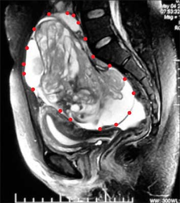
Fig. 1 MRI abdomen pelvis showing complex right ovarian mass, marked by red dots.
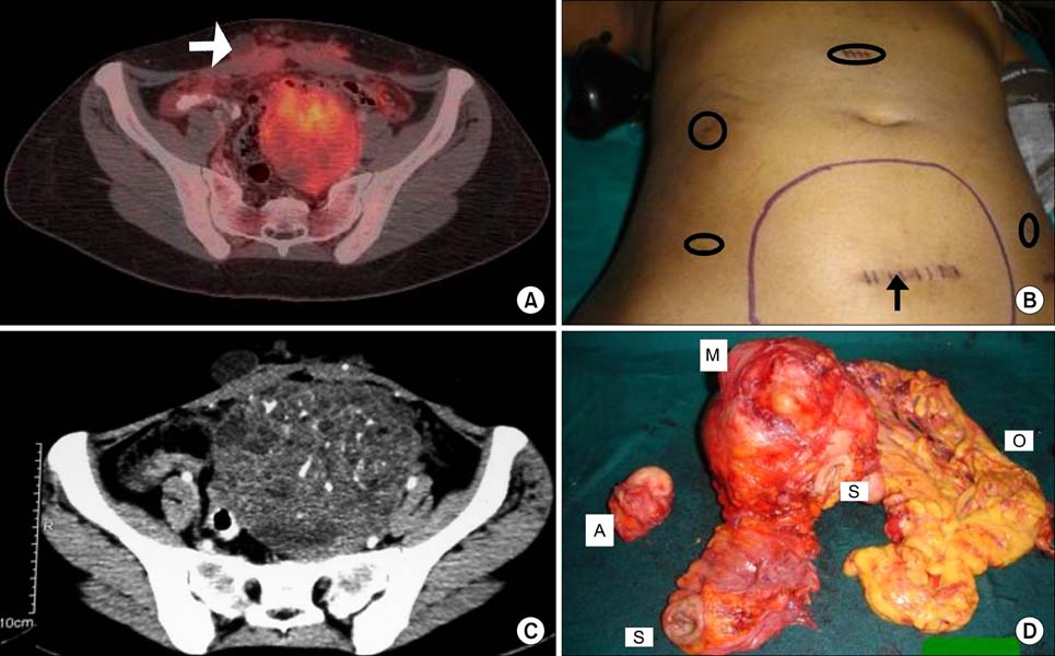
Fig. 2 (A) PET-CT pelvis showing fluoro-2-deoxy-D-glucose (FDG) avid pelvic mass and abdominal wall deposit (marked by white arrow). (B) Photograph showing abdomino-pelvic mass marked in blue. Black arrow shows abdominal wall mass (port site of specimen extraction), other ports marked by black ellipse. (C) CT scan abdomen pelvis post chemotherapy showing increased calcification in pelvic mass. (D) Specimen showing pelvic mass (M), two ends of sigmoid colon (S), anterior abdominal wall deposit (A), and omentum (O).
Reference
-
1. DiSaia PJ, Saltz A, Kagan AR, Morrow CP. Chemotherapeutic retroconversion of immature teratoma of the ovary. Obstet Gynecol. 1977. 49:346–350.2. Logothetis CJ, Samuels ML, Trindade A, Johnson DE. The growing teratoma syndrome. Cancer. 1982. 50:1629–1635.3. Moskovic E, Jobling T, Fisher C, Wiltshaw E, Parsons C. Retroconversion of immature teratoma of the ovary: CT appearances. Clin Radiol. 1991. 43:402–408.4. Andre F, Fizazi K, Culine S, Droz J, Taupin P, Lhomme C, et al. The growing teratoma syndrome: results of therapy and long term follow-up of 33 patients. Eur J Cancer. 2000. 36:1389–1394.5. Dixon FJ, Moore RA. Armed Forces Institute of Pathology. Tumors of the male sex organs. Atlas of tumor pathology. 1952. Vol 8. Washington, DC: Armed Forces Institute of Pathology;316–332.6. Williams SD, Blessing JA, DiSaia PJ, Major FJ, Ball HG 3rd, Liao SY. Second-look laparotomy in ovarian germ cell tumors: the Gynecologic Oncology Group experience. Gynecol Oncol. 1994. 52:287–291.7. Hariprasad R, Kumar L, Janga D, Kumar S, Vijayaraghavan M. Growing teratoma syndrome of ovary. Int J Clin Oncol. 2008. 13:83–87.8. Jumean HG, Komorowski R, Mahvi D, Anderson T. Immature teratoma of the ovary: an unusual case. Gynecol Oncol. 1992. 46:111–114.9. Kattan J, Droz JP, Culine S, Duvillard P, Thiellet A, Peillon C. The growing teratoma syndrome: a woman with nonseminomatous germ cell tumor of ovary. Gynecol Oncol. 1993. 49:395–399.10. Nimkin K, Gupta P, McCauley R, Gilchrist BF, Lessin MS. The growing teratoma syndrome. Paediatr Radiol. 2004. 34:259–262.
- Full Text Links
-
- Actions
-
Cited
- CITED
-
- Close
- Share
-
- Similar articles
-
- A Case of Recurrent Growing Teratoma Syndrome during Chemotherapy for Immature Teratoma of the Ovary
- A case of congenital retroperitoneal immature teratoma
- Low-grade Immature Teratoma of the Ovary with Gliomatosis Peritonei: A case report
- A case of sequent occurrence of mature and immature teratomas in same patient
- A Case of Gliomatosis Peritonei
