Bizarre Parosteal Osteochondromatous Proliferation with Cortical Erosion in the Hand: A Case Report
- Affiliations
-
- 1Department of Radiology, Inje University College of Medicine, Ilsan Paik Hospital, Goyang, Korea. kyh@paik.ac.kr
- KMID: 2002945
- DOI: http://doi.org/10.3348/jksr.2011.65.4.415
Abstract
- Bizarre parosteal osteochondromatous proliferation (BPOP) is a rare osseous lesion occurring in the long bones of distal extremities. Typical imaging findings of BPOP show a well-defined calcified osseous mass without soft tissue swelling, cortical destruction, and medullary involvement. We experienced rare aggressive magnetic resonance imaging findings of BPOP with cortical erosion, bone marrow edema, and adjacent soft tissue enhancement, which was misdiagnosed as a malignancy or inflammatory lesion.
Figure
-

Fig. 1 AP (A) and lateral (B) plain radiographs show a well-defined exophytic osseous mass arising from volar aspect of the right 2nd proximal phalanx shaft. The mass has a sessile appearance to underlying bone with sclerosis.

Fig. 2 MR images of BPOP. Axial T1-weighted image (TR/TE 540/15) (A) shows iso-signal intensity with muscle and overlying cortical erosion. On a coronal T2-weighted image (TR/TE 2700/96) (B), the mass reveals bright high signal intensity (arrow). Also, high signal intensity is noted in adjacent soft tissue and bone marrow, suggesting edema. Contrast-enhanced fat suppression axial (TR/TE 420/15) (C) and sagittal (TR/TE 510/20) (D) T1-weighted images show homogeneous well enhancement of the mass. Surrounding soft tissue and bone marrow shows enhancement. Peripheral low signal intensity rim of mass on T1 and T2 weighted images corresponds to a sclerotic rim on simple radiograph. Note.-BPOP = bizarre parosteal osteochondromatous proliferation
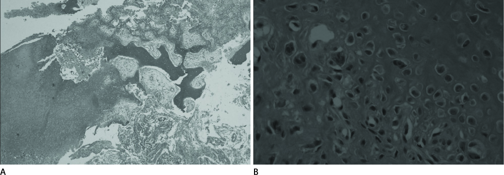
Fig. 3 Upon microscopic examination, the mass shows a mixture of hypercellular cartilage maturing to trabecular bone with a classic irregular chondro-osseous interface. Fragments of fasciitis-like stromal proliferation, proliferative cartilage, as well as woven and cortical-like bone formation with rims of osteoblast are noted in the resected mass (H&E stain, × 40) (A). Irregular maturation of cartilage with hypercellular, occasionally binucleated, enlarged chondrocytes is noted in the mass (H&E stain, × 200) (B).
Cited by 1 articles
-
Diagnosis and Treatment of Bizarre Parosteal Osteochondromatous Proliferation
Duk-Seop Shin, Han Seok Yang, Ho Dong Na, Seung Min Ryu
J Korean Orthop Assoc. 2018;53(1):58-65. doi: 10.4055/jkoa.2018.53.1.58.
Reference
-
1. Torreggiani WC, Munk PL, Al-Ismail K, O'Connell JX, Nicolaou S, Lee MJ, et al. MR imaging features of bizarre parosteal osteochondromatous proliferation of bone (Nora's lesion). Eur J Radiol. 2001; 40:224–231.2. Soon JL, Chang HC, Sim CS, Teoh LC, Low CO. A case of Bizarre Parosteal Osteochondromatous Proliferation of the hand. Singapore Med J. 2003; 44:27–30.3. Joseph J, Ritchie D, MacDuff E, Mahendra A. Bizarre parosteal osteochondromatous proliferation: a locally aggressive benign tumor. Clin Orthop Relat Res. 2011; 469:2019–2027.4. Murphey MD, Choi JJ, Kransdorf MJ, Flemming DJ, Gannon FH. Imaging of osteochondroma: variants and complications with radiologic-pathologic correlation. Radiographics. 2000; 20:1407–1434.5. Helliwell TR, O'Connor MA, Ritchie DA, Feldberg L, Stilwell JH, Jane MJ. Bizarre parosteal osteochondromatous proliferation with cortical invasion. Skeletal Radiol. 2001; 30:282–285.6. Ly JQ, Bui-Mansfield LT, Taylor DC. Radiologic demonstration of temporal development of bizarre parosteal osteochondromatous proliferation. Clin Imaging. 2004; 28:216–218.7. Meneses MF, Unni KK, Swee RG. Bizarre parosteal osteochondromatous proliferation of bone (Nora's lesion). Am J Surg Pathol. 1993; 17:691–697.8. Yuen M, Friedman L, Orr W, Cockshott WP. Proliferative periosteal processes of phalanges: a unitary hypothesis. Skeletal Radiol. 1992; 21:301–303.9. Sakamoto A, Imamura S, Matsumoto Y, Harimaya K, Matsuda S, Takahashi Y, et al. Bizarre parosteal osteochondromatous proliferation with an inversion of chromosome 7. Skeletal Radiol. 2011; 40:1487–1490.10. Choi JH, Gu MJ, Kim MJ, Choi WH, Shin DS, Cho KH. Fibrosarcoma in bizarre parosteal osteochondromatous proliferation. Skeletal Radiol. 2001; 30:44–47.11. Michelsen H, Abramovici L, Steiner G, Posner MA. Bizarre parosteal osteochondromatous proliferation (Nora's lesion) in the hand. J Hand Surg Am. 2004; 29:520–525.
- Full Text Links
-
- Actions
-
Cited
- CITED
-
- Close
- Share
-
- Similar articles
-
- Parosteal Lipoma Associated with Underlying Recurrent Bizarre Parosteal Osteochondromatous Proliferation (Nora's Lesion) of the Hand
- Bizarre Parosteal Osteochondromatous Proliferation of Middle Phalanx: A Case Report
- A Case of Subungual Bizarre Parosteal Osteochondromatous Proliferation of the Toe
- Bizarre Parosteal Osteochondromatous Proliferation: A Report of One Case
- Aberrant Chromosomal Alterations in Bizarre Parosteal Osteochondromatous Proliferation (Nora's Lesion) of Hand: A Case Report
