Splenogonadal Fusion: A Case Report
- Affiliations
-
- 1Department of Radiology, Wonju Christian Hospital, Yonsei University Wonju College of Medicine, Wonju, Korea. wckwon@yonsei.ac.kr
- KMID: 2002927
- DOI: http://doi.org/10.3348/jksr.2011.65.6.589
Abstract
- Splenogonadal fusion is a rare congenital anomaly characaterized as the fusion of the spleen and a gonad. The first report of this anomaly was in 1883 and only 160 cases have been reported to date. As far as we know, this is the first reported case of splenogonadal fusion in this country.
MeSH Terms
Figure
-

Fig. 1 Scrotal ultrasound. A. A longitudinal grayscale scan shows an oval-shaped extratesticular mass (arrow) and a downward displaced left testis (open arrow). B. Power Doppler shows increased vascularity of the extratesticular mass compared with the testis. C. Note the normal right testis. Left testis shows a normal size and shape with no connection between the left testis and mass. D. Power Doppler also shows multiple engorged vessels surrounding the extratesticular mass.
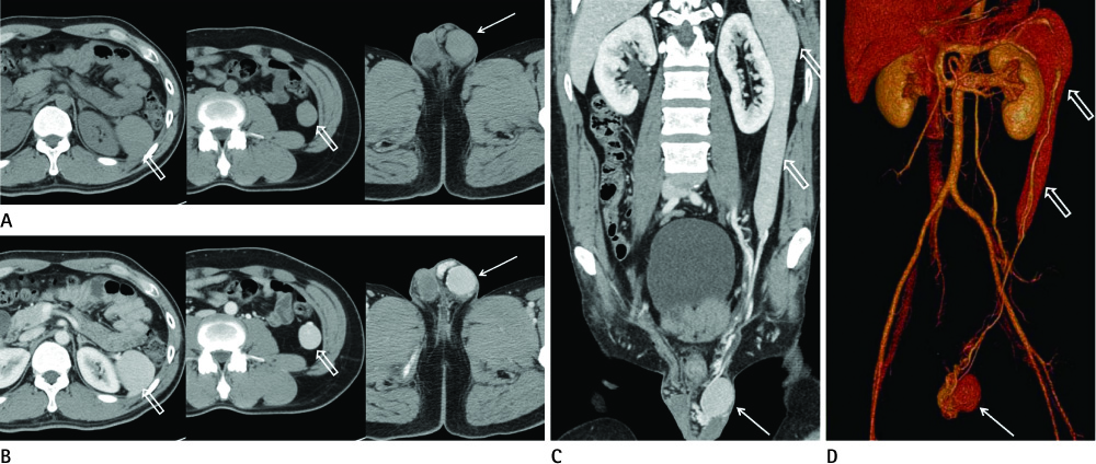
Fig. 2 2. Abdominal CT scan. A. An unenhanced scan of the left scrotal mass (arrow) shows homogeneous density. B. An axial contrast-enhanced CT scan shows a left extratesticular mass (arrow) homogeneously enhanced to a similar degree to that of the spleen (open arrow). C, D. A multi-planar reconstruction image (C) and a three-dimensional volume rendering image (D) shows an elongated spleen (open arrows) and left scrotal mass (arrow) which are connected to each other by engorged splenic vessels.

Fig. 3 Abdominal MRI. A, B. Axial T1 weighted image (WI) (left) and T2WI (right) shows a left extratesticular mass (arrow) with similar signal intensity of the spleen (open arrow). C. Curved coronal T2WI shows an elongated spleen (open arrow) and left scrotal mass (arrow) which are connected by the splenic vessels.

Fig. 4 A red blood cell scan using 99m Tc showing an elongated spleen with a tail-like projection to the left pelvic cavity (arrow) and oval-shaped increased radioactivity in left scrotum which is connected with the spleen through band-like radioactivity (open arrow). Radioactivity of the left scrotum may be more intense than that of the spleen because of the composite activity of the engorged left scrotal vessels. Engorged left scrotal vessels can be also shown on ultrasonogram (Fig. 1D).
Reference
-
1. Ando S, Shimazui T, Hattori K, Yamamoto T, Kuriyagawa K, Akaza H. Splenogonadal fusion: case report and review of published works. Int J Urol. 2006; 13:1539–1541.2. Alvarez Maestro M, López-Tello J, Domínguez Franjo P, Ríos González E, Martínez-Piñeiro L. [Splenogonadal fusion. Report of a case and review of the literature]. Actas Urol Esp. 2010; 34:293–295.3. Boestrom E. Demonstration eines praparates von verwachsung der milz mit dem linken hoden. Gellschaft deutscher Naturforscher und Artze Verhandlungen der 56 Versammlung. Freiburg. 1883; 149.4. Putschar WG, Manion WC. Splenicgonadal fusion. Am J Pathol. 1956; 32:15–33.5. Carragher AM. One hundred years of splenogonadal fusion. Urology. 1990; 35:471–475.6. Duncan WL Jr, Barraza MA. Splenogonadal fusion: a case report and review of literature. J Pediatr Surg. 2005; 40:e5–e7.7. Hizli F, Uygur MC, Irkkan C. Splenogonadal fusion: report of a case. Int J Urol. 2005; 12:591–592.8. Sneath WA. An apparent third testicle consisting of a scrotal spleen. J Anat Physiol. 1913; 47:340–342.9. Steinmetz AP, Rappaport A, Nikolov G, Priel IE, Chamovitz DL, Dolev E. Splenogonadal fusion diagnosed by spleen scintigraphy. J Nucl Med. 1997; 38:1153–1155.
- Full Text Links
-
- Actions
-
Cited
- CITED
-
- Close
- Share
-
- Similar articles
-
- Intertransverse Fusion in Spondylolisthesis: Report of a Case
- Repeated Migration of a Fusion Cage after Posterior Lumbar Interbody Fusion
- Sacral Stress Fracture Developing after Lumbosacral Fusion in a Patient with Spondylolisthesis: A Case Report
- The Treatment of Spondylolysis with Anterior Spinal Fusion: A Report of Three Cases
- Severe Retrolisthesis at the Adjacent Segment after Lumbar Fusion Combined with Dynamic Stabilization
