Testicular Epidermoid Cyst on Diffusion-Weighted MR Imaging and ADC Map : A Case Report
- Affiliations
-
- 1Department of Radiology, Ilsan Paik Hospital, Inje University College of Medicine, Korea.
- 2Department of Radiology, Yonsei University College of Medicine, Korea. bellenina@daum.net
- 3Department of Pathology, Ilsan Paik Hospital, Inje University College of Medicine, Korea.
- KMID: 2000064
- DOI: http://doi.org/10.13104/jksmrm.2011.15.2.154
Abstract
- Testicular epidermoid cyst is a rare benign tumor, accounting for 1-2% of all testicular tumors. It can be cured by organ preserving surgery, so accurate preoperative diagnosis is very important for preventing unneccessary and extensive orchiectomy. We experienced a case of an 18-year-old man who presented with a painless lump in his right testis. The testicular mass showed an onion ring sign on ultrasonography. Computed tomography images showed the mass as a low attenuating lesion with curvilinear calcification. On Magnetic resonance imaging (MRI), the mass appeared as high signal intensity with internal alternating low signal intensity patterns on T2-weighted images. The mass was displayed as having homogeneous high signal intensity on diffusion magnetic resonance imaging and showed lower apparent diffusion coefficient values than normal testis parenchyma, similar to intracranial epidermoid cysts. Testicular MRI with DWI and ADC map can help to more accurately diagnose testicular epidermoid cyst.
MeSH Terms
Figure
-
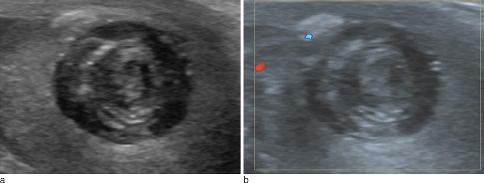
Fig. 1 Ultrasonography (US) finding for the testicular epidermoid cyst. (a) Gray scale transverse US reveals a well-circumscribed intratesticular mass in the mid portion of right testis. It shows alternating rings of hyper- and hypoechogenecities. (b) Doppler US reveals no vascularity in the mass.

Fig. 2 CT shows a well-defined non-enhancing cystic mass in the right testis. (a) Non-enhanced axial CT image reveals focal curvilinear calcification (arrow). (b) Enhanced axial CT image shows no enhancement of the mass.
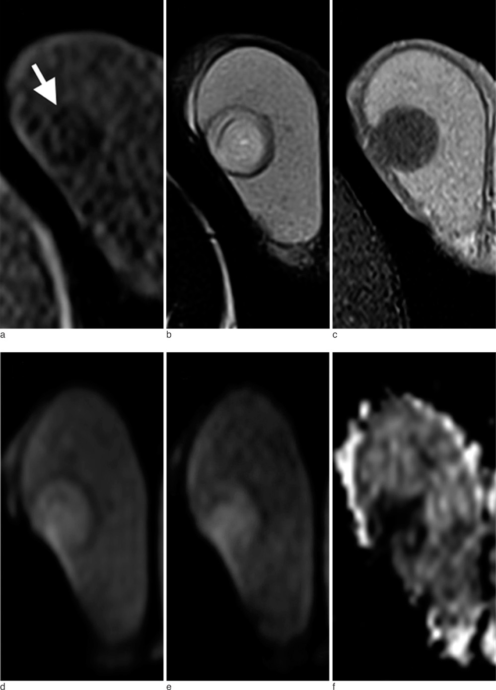
Fig. 3 MR images of the testicular epidermoid cyst. (a) Axial T1-weighted image (TR/TE 90/4.9) shows a well-circumscribed low signal intensity mass with a low signal rim (arrow). (b) Axial T2-weighted image (TR/TE 3000/92) shows high signal intensity with multi-layered concentric low signal rings. Peripheral low signal rim is seen on both T1-weighted image and T2-weighted image. (c) Axial gadolinium-enhanced T1-weighted fat suppression image (TR/TE 724/9.6) shows no contrast enhancement. (d, e) DWI shows higher signal intensity in the tumor than in the adjacent normal testicular parenchyma (b-value 50, 800). (f) ADC map shows lower ADC values than in the normal testicular parenchyma.

Fig. 4 (a) Gross specimen reveals a well-encapsulated whitish mass of 2×2 cm in size. (b) Upon microscopic examination, the wall of the testicular mass is lined by squamous epithelium (arrowhead) with a granular cell layer and a fibrous wall (arrow) of variable thickness, and it is filled with keratin.
Reference
-
1. Cho JH, Chang JC, Park BH, Lee JG, Son CH. Sonographic and MR imaging findings of testicular epidermoid cysts. AJR Am J Roentgenol. 2002. 178:743–748.2. Saremi F, Knoll AN, Bendavid OJ, Schultze-Haakh H, Narula N, Sarlati F. Characterization of genitourinary lesions with diffusion-weighted imaging. Radiographics. 2009. 29:1295–1317.3. Hakyemez B, Aksoy U, Yildiz H, Ergin N. Intracranial epidermoid cysts: diffusion-weighted, FLAIR and conventional MR findings. Eur J Radiol. 2005. 54:214–220.4. Atchley JT, Dewbury KC. Ultrasound appearances of testicular epidermoid cysts. Clin Radiol. 2000. 55:493–502.5. Langer JE, Ramchandani P, Siegelman ES, Banner MP. Epidermoid cysts of the testicle: sonographic and MR imaging features. AJR Am J Roentgenol. 1999. 173:1295–1299.6. Stein MM, Stein MW, Cohen BC, Li M, Koenigsberg M. Unusual sonographic appearance of an epidermoid cyst of the testis. J Ultrasound Med. 1999. 18:723–726.7. Maizlin ZV, Belenky A, Baniel J, Gottlieb P, Sandbank J, Strauss S. Epidermoid cyst and teratoma of the testis: sonographic and histologic similarities. J Ultrasound Med. 2005. 24:1403–1409. quiz 1410–quiz 1401.8. Chen C, Wu C, LIN W, Cheng W, Sun G, Chen C. Unusual MR imagings of an incidental testicular epidermoid Cyst: a case report. Chin J Radiol. 2005. 30:251–256.9. Woodward PJ, Sohaey R, O'Donoghue MJ, Green DE. From the archives of the AFIP: tumors and tumorlike lesions of the testis: radiologic-pathologic correlation. Radiographics. 2002. 22:189–216.10. Yoshida T. MRI of testicular epidermoid cyst. Radiat Med. 2004. 22:354–356.
- Full Text Links
-
- Actions
-
Cited
- CITED
-
- Close
- Share
-
- Similar articles
-
- The Usefulness of Diffusion Weighted Imaging in the Differential Diagnosis of Various Intracranial Cystic Lesions
- Clinical applications and characteristics of apparent diffusion coefficient maps for the brain of two dogs
- Epidermoid Cyst of the Testis Demonstrating Double Target Sign: A Case Report
- SNR and ADC Changes at Increasing b Values among Patients with Lumbar Vertebral Compression Fracture on 1.5T MR Diffusion Weighted Images
- Diffusion-Weighted MR Imaging of Unicystic Odontogenic Tumors for Differentiation of Unicystic Ameloblastomas from Keratocystic Odontogenic Tumors
