Combined small cell carcinoma and adenocarcinoma arising in uterine cervix
- Affiliations
-
- 1Department of Pathology, Yangji General Hospital, Seoul, Korea. laghonda@naver.com
- 2Department of Gynecology, Cheju Halla General Hospital, Jeju, Korea.
- 3Department of Radiology, Cheju Halla General Hospital, Jeju, Korea.
- KMID: 1992625
- DOI: http://doi.org/10.5468/KJOG.2012.55.4.269
Abstract
- We report here on a case of combined small cell carcinoma and adenocarcinoma arising on uterine cervix. The patient was a 39-year-old woman who presented with uterine cervical mass. Colposcopic biopsy confirmed as adenocarcinoma and radical hysterectomy was performed. The mass, measuring in about 2 cm in greatest diameter, was mainly endophytic growing. Histopathologically, the mass was consisted of well differentiated adenocarcinoma from 2 to 9 o'clock directions of uterine cervix. From 9 to 11 o'clock directions, small cell carcinoma was detected. On results of special stain and immunohistochemistry, mucin stains and carcinoembryonic antigen were positive in area of adenocarcinoma. Chromogranin A and synaptophysin, stain for neuroendocrine markers, were positive in region of small cell carcinoma. This case indicates that the small cell carcinoma and adenocarcinoma can simultaneously occur in uterine cervix.
Keyword
MeSH Terms
Figure
-
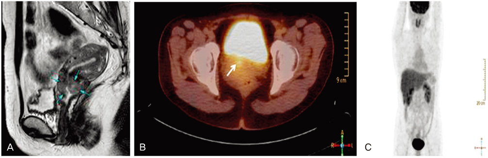
Fig. 1 Radiologic findings of the cervical tumor. (A) Pelvic magnetic resonance imaging shows the mass with increased signal intensity in the endocervical area of uterus. (B, C) Positron emission tomography (PET)/computerized tomography images show a hypermetabolic focus on lower uterine cervix. Note no evidence of distant tumor metastasis on PET scan.
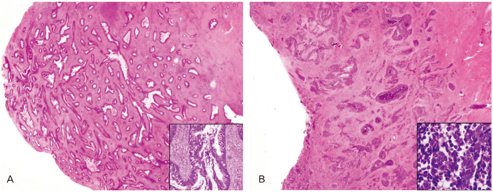
Fig. 2 (A) On microscopic findings, the mounted section shows that well differentiated adenocarcinoma of the cervix infiltrating the stroma. The inset shows pseudostratified nuclei and cribriform pattern (A; H&E, ×200). (B) Small cell carcinoma shows confluent sheets of darkly staining neoplastic cells infiltrate the stroma on mounted section. The inset shows the loose packed tumor cells with scant cytoplasm and small foci of necrosis (B; H&E, ×200).

Fig. 3 Special stains and immunohistochemical stainings. (A) Adenocarcinoma (Periodic acid-schiff stain, ×400). (B) Carcinoembryonic antigen in adenocarcinoma (×400). (C) Chromogranin A in small cell carcinoma (×400). (D) Synaptophysin in small cell carcinoma (×400).
Reference
-
1. Bermúdez A, Vighi S, García A, Sardi J. Neuroendocrine cervical carcinoma: a diagnostic and therapeutic challenge. Gynecol Oncol. 2001. 82:32–39.2. Mannion C, Park WS, Man YG, Zhuang Z, Albores-Saavedra J, Tavassoli FA. Endocrine tumors of the cervix: morphologic assessment, expression of human papillomavirus, and evaluation for loss of heterozygosity on 1p,3p, 11q, and 17p. Cancer. 1998. 83:1391–1400.3. Seong HJ, Kim JY, Lee SM, Do YR, Song HS. A case of small cell carcinoma of the uterine cervix. Korean J Med. 2008. 74:208–213.4. Albores-Saavedra J, Larrazag O, Poucell S, Rodriguez-Martinez HA. Primary carcinoid of the uterine cervix. Patologia. 1972. 10:185–193.5. Sheets EE, Berman ML, Hrountas CK, Liao SY, DiSaia PJ. Surgically treated, early-stage neuroendocrine small-cell cervical carcinoma. Obstet Gynecol. 1988. 71:10–14.6. Straughn JM Jr, Richter HE, Conner MG, Meleth S, Barnes MN. Predictors of outcome in small cell carcinoma of the cervix: a case series. Gynecol Oncol. 2001. 83:216–220.7. Masumoto N, Fujii T, Ishikawa M, Saito M, Iwata T, Fukuchi T, et al. P16 overexpression and human papillomavirus infection in small cell carcinoma of the uterine cervix. Hum Pathol. 2003. 34:778–783.8. Gilks CB, Young RH, Gersell DJ, Clement PB. Large cell neuroendocrine [corrected] carcinoma of the uterine cervix: a clinicopathologic study of 12 cases. Am J Surg Pathol. 1997. 21:905–914.9. Horn LC, Hentschel B, Bilek K, Richter CE, Einenkel J, Leo C. Mixed small cell carcinomas of the uterine cervix: prognostic impact of focal neuroendocrine differentiation but not of Ki-67 labeling index. Ann Diagn Pathol. 2006. 10:140–143.10. Lancaster WD, Castellano C, Santos C, Delgado G, Kurman RJ, Jenson AB. Human papillomavirus deoxyribonucleic acid in cervical carcinoma from primary and metastatic sites. Am J Obstet Gynecol. 1986. 154:115–119.11. Groben P, Reddick R, Askin F. The pathologic spectrum of small cell carcinoma of the cervix. Int J Gynecol Pathol. 1985. 4:42–57.12. Gersell DJ, Mazoujian G, Mutch DG, Rudloff MA. Small-cell undifferentiated carcinoma of the cervix. A clinicopathologic, ultrastructural, and immunocytochemical study of 15 cases. Am J Surg Pathol. 1988. 12:684–698.13. Dabbs DJ. Diagnostic immunohistochemistry. 2006. 2nd ed. Edinburgh: Churchill Livingstone.14. Cohen JG, Kapp DS, Shin JY, Urban R, Sherman AE, Chen LM, et al. Small cell carcinoma of the cervix: treatment and survival outcomes of 188 patients. Am J Obstet Gynecol. 2010. 203:347.e1-6.
- Full Text Links
-
- Actions
-
Cited
- CITED
-
- Close
- Share
-
- Similar articles
-
- A case of small cell carcinoma of the uterine cervix
- Small Cell Carcinoma of the Uterine Cervix: A Clinicophthologic, Ultrastructural, and Immunohistochemical Study of 4 Cases
- Endometrioid Adenocarcinoma Arising from Endometriosis of the Uterine Cervix: A Case Report
- A case of clear cell carcinoma that is unrelated to diethystilbestrol of the uterine cervix
- Small cell neuroendocrine carcinoma of the uterine cervix presenting with syndrome of inappropriate antidiuretic hormone secretion
