Prevention of anti-SSA/Ro and anti-SSB/La antibodies-mediated congenital heart block in pregnant woman with systemic lupus erythematosus: A case report
- Affiliations
-
- 1Department of Obstetrics and Gynecology, Konyang University College of Medicine, Daejeon, Korea. sungeunog@hanmail.net
- KMID: 1992560
- DOI: http://doi.org/10.5468/KJOG.2012.55.7.502
Abstract
- Women with connective tissue diseases increase risks of obstetric complications during pregnancy. Especially, women who have anti-SSA/Ro and anti-SSB/La auto-antibodies increase the risk of developing fetal atrioventricular (AV) block. It is difficult to indentify fetal AV block in the early stage of the disease, which is consequently found after the establishment of complete AV block as the form of fetal arrhythmia. To have a chance to reverse this serious disorder, we should detect fetal AV block in the early stage before it progresses to complete AV block. We experienced a case of pregnant systemic lupus erythematosus woman who has anti-SSA/Ro and anti-SSB/La auto-antibodies monitored by cardiac doppler sonography; We detected fetal first degree AV block and successfully treated with dexamethasone. We report systemic lupus erythematosus women who increased risks of fetal AV block with a brief review of relevant literatures.
Keyword
MeSH Terms
Figure
-
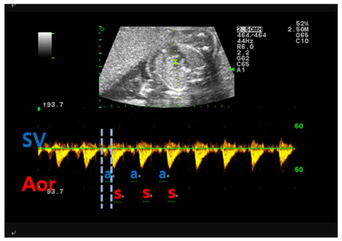
Fig. 1 Pulsed Doppler flow curves showing normal atrioventricular relationship of cardiac rhythm. Doppler tracing from the superior vena cava (SVC) and ascending aorta. SVC flow has small atrial reversed wave (a). The aortic flow curve consists of a forward peak wave (s). Atrioventricular time interval is the time lapse between the onsets of vena caval a-wave and aortic s-wave (between grey lines).
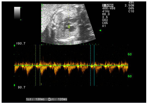
Fig. 2 Pulsed Doppler was done at 26+1 weeks of gestation. Checked atrioventricular time intervals were 138 ms and 120 ms. These values were prolonged compared to normal range (reference range, 111±17 ms).
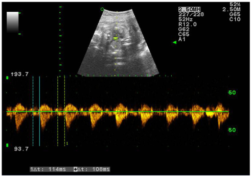
Fig. 3 Pulsed Doppler was done at 27+1 weeks of gestation. Checked atrioventricular time intervals were 114 ms and 108 ms. These values were within the normal range (reference range, 111±17 ms).
Cited by 1 articles
-
Fetal Congenital Complete Atrioventricular Block in a Mother with Isolated Serum Anti-SSA/Ro and Anti-SSB/La Antibodies
Chi-Won Mok, Ju-Young Park, Kyunghee Kim, Suk-Joo Choi, Soo-young Oh, Cheong-Rae Roh, Jong-Hwa Kim
Perinatology. 2016;27(3):185-189. doi: 10.14734/PN.2016.27.3.185.
Reference
-
1. Buyon JP, Hiebert R, Copel J, Craft J, Friedman D, Katholi M, et al. Autoimmune-associated congenital heart block: demographics, mortality, morbidity and recurrence rates obtained from a national neonatal lupus registry. J Am Coll Cardiol. 1998. 31:1658–1666.2. Fouron JC, Proulx F, Miró J, Gosselin J. Doppler and M-mode ultrasonography to time fetal atrial and ventricular contractions. Obstet Gynecol. 2000. 96:732–736.3. D'Cruz DP, Khamashta MA, Hughes GR. Systemic lupus erythematosus. Lancet. 2007. 369:587–596.4. Mok CC, Wong RW. Pregnancy in systemic lupus erythematosus. Postgrad Med J. 2001. 77:157–165.5. Vesel S, Mazic U, Blejec T, Podnar T. First-degree heart block in the fetus of an anti-SSA/Ro-positive mother: reversal after a short course of dexamethasone treatment. Arthritis Rheum. 2004. 50:2223–2226.6. Rein AJ, Mevorach D, Perles Z, Gavri S, Nadjari M, Nir A, et al. Early diagnosis and treatment of atrioventricular block in the fetus exposed to maternal anti-SSA/Ro-SSB/La antibodies: a prospective, observational, fetal kinetocardiogram-based study. Circulation. 2009. 119:1867–1872.7. Waltuck J, Buyon JP. Autoantibody-associated congenital heart block: outcome in mothers and children. Ann Intern Med. 1994. 120:544–551.8. Moak JP, Barron KS, Hougen TJ, Wiles HB, Balaji S, Sreeram N, et al. Congenital heart block: development of late-onset cardiomyopathy, a previously underappreciated sequela. J Am Coll Cardiol. 2001. 37:238–242.9. Saleeb S, Copel J, Friedman D, Buyon JP. Comparison of treatment with fluorinated glucocorticoids to the natural history of autoantibody-associated congenital heart block: retrospective review of the research registry for neonatal lupus. Arthritis Rheum. 1999. 42:2335–2345.10. Brucato A. Prevention of congenital heart block in children of SSA-positive mothers. Rheumatology (Oxford). 2008. 47:Suppl 3. iii35–iii37.11. Ostensen M, Khamashta M, Lockshin M, Parke A, Brucato A, Carp H, et al. Anti-inflammatory and immunosuppressive drugs and reproduction. Arthritis Res Ther. 2006. 8:209.12. Lee BH, Stoll BJ, McDonald SA, Higgins RD. National Institute of Child Health and Human Development Neonatal Research Network. Neurodevelopmental outcomes of extremely low birth weight infants exposed prenatally to dexamethasone versus betamethasone. Pediatrics. 2008. 121:289–296.13. Brownfoot FC, Crowther CA, Middleton P. Different corticosteroids and regimens for accelerating fetal lung maturation for women at risk of preterm birth. Cochrane Database Syst Rev. 2008. (4):CD006764.14. Chung SH, Lee Y, Cheon YH, Jung IC, Yoon WS, Lee JS, et al. A case of neonatal lupus syndrome with congenital complete heart block. Korean J Obstet Gynecol. 2002. 45:723–727.15. Sonesson SE, Salomonsson S, Jacobsson LA, Bremme K, Wahren-Herlenius M. Signs of first-degree heart block occur in one-third of fetuses of pregnant women with anti-SSA/Ro 52-kd antibodies. Arthritis Rheum. 2004. 50:1253–1261.
