Korean J Urol.
2012 Dec;53(12):875-878. 10.4111/kju.2012.53.12.875.
A Case of Renal Schwannoma
- Affiliations
-
- 1Department of Urology, Soonchunhyang University College of Medicine, Cheonan, Korea. ysurol@schmc.ac.kr
- 2Department of Pathology, Soonchunhyang University College of Medicine, Cheonan, Korea.
- KMID: 1988735
- DOI: http://doi.org/10.4111/kju.2012.53.12.875
Abstract
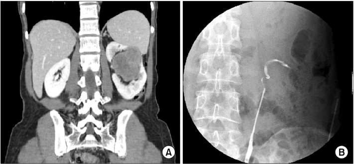
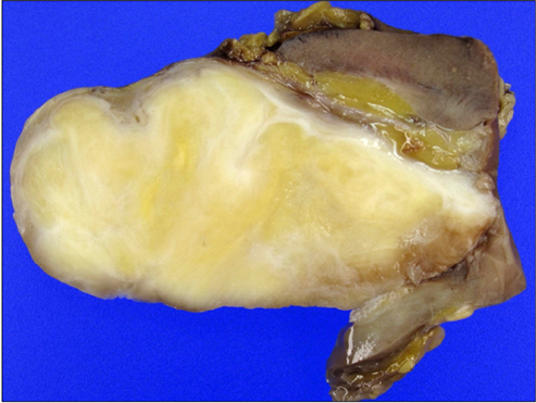
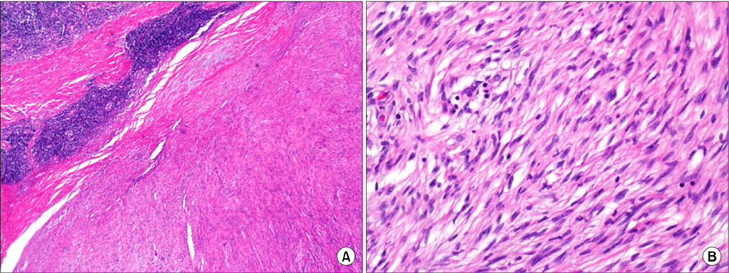
- Schwannomas are benign tumors that arise from the neural sheath of Schwann cells. Renal schwannomas are extremely rare and are commonly misdiagnosed as renal cell carcinoma, which typically results in a radical nephrectomy. We present a case of a renal schwannoma that mimics a renal pelvis tumor.
Keyword
Figure
Cited by 1 articles
-
Radiologic Findings of Renal Schwannoma: A Case Report and Literature Review
Sung Tae Hwang, Deuk Jae Sung, Ki Choon Sim, Na Yeon Han, Beom Jin Park, Min Ju Kim, Jeong Hyeon Lee
J Korean Soc Radiol. 2018;78(4):289-294. doi: 10.3348/jksr.2018.78.4.289.
Reference
-
1. Gubbay AD, Moschilla G, Gray BN, Thompson I. Retroperitoneal schwannoma: a case series and review. Aust N Z J Surg. 1995. 65:197–200.2. Hung SF, Chung SD, Lai MK, Chueh SC, Yu HJ. Renal Schwannoma: case report and literature review. Urology. 2008. 72:716.e3–716.e6.3. Gobbo S, Eble JN, Huang J, Grignon DJ, Wang M, Martignoni G, et al. Schwannoma of the kidney. Mod Pathol. 2008. 21:779–783.4. Ghiatas AA, Faleski EJ. Benign solitary schwannoma of the retroperitoneum: CT features. South Med J. 1989. 82:801–802.5. Kitagawa K, Yamahana T, Hirano S, Kawaguchi S, Mikawa I, Masuda S, et al. MR imaging of neurilemoma arising from the renal hilus. J Comput Assist Tomogr. 1990. 14:830–832.6. Daneshmand S, Youssefzadeh D, Chamie K, Boswell W, Wu N, Stein JP, et al. Benign retroperitoneal schwannoma: a case series and review of the literature. Urology. 2003. 62:993–997.7. Alvarado-Cabrero I, Folpe AL, Srigley JR, Gaudin P, Philip AT, Reuter VE, et al. Intrarenal schwannoma: a report of four cases including three cellular variants. Mod Pathol. 2000. 13:851–856.8. Nishio A, Adachi W, Igarashi J, Koide N, Kajikawa S, Amano J. Laparoscopic resection of a retroperitoneal schwannoma. Surg Laparosc Endosc Percutan Tech. 1999. 9:306–309.
- Full Text Links
- Actions
-
Cited
- CITED
-
- Close
- Share
- Similar articles
-
- Radiologic Findings of Renal Schwannoma: A Case Report and Literature Review
- Hypervascular Vestibular Schwannoma: A Case Report
- A Case of Schwannoma of the Chorda Tympani Nerve
- Melanotic Schwannoma in Cervical Spine: A Case Report
- A Case of Pelvic Retroperitoneal Schwannoma : Preoperatively Suspected of Malignant Adnexal Tumor


