Noncompliance screw supported maxillary molar distalization in a parallel manner
- Affiliations
-
- 1Department of Orthodontics, Faculty of Dentistry, Cumhuriyet University, Sivas, Turkey. altugbicakci@gmail.com
- 2Ankara Mevki Military Hospital, Ankara, Turkey.
- KMID: 1975605
- DOI: http://doi.org/10.4041/kjod.2010.40.4.250
Abstract
OBJECTIVE
Intraoral noncompliance upper molar distalization techniques have gained in popularity and have subsequently found to be successful in Class II correction. The aim of the present study was to introduce a screw supported intraoral distalization appliance and investigate its efficiency.
METHODS
Twenty-one subjects (11 females, 10 males; average age of 14.9 years) with Angle Class II malocclusion participated in this study. Two screws were inserted behind the incisive foramen and immediately loaded to distalize the upper first molars. An intraoral screw supported distalization appliance was used to distalize the upper molars in order to achieve a Class I molar relationship. Skeletal and dental changes were evaluated using cephalometric and three-dimensional (3D) model analysis.
RESULTS
Upper molars were distalized 3.95 mm on average and a Class I molar relationship was achieved without any anchorage loss. The upper molars were tipped only 1.49degrees and the upper right and left molars were rotated only 0.54degrees and 0.74degrees respectively which were statistically non-significant (p > 0.05).
CONCLUSIONS
The newly designed screw supported noncompliance distalization appliance was found to be an effective device for achieving bodily molar distalization without any anchorage loss.
Figure
-

Fig. 1 Fabrication of screw supported distalization appliance. 'U' bending of 0.040 inch stainless steel rod at the level of distal surface of the second molar was performed and extended into the acrylic part of the appliance to enhance its stability. A superelastic heavy open-coil spring is compressed along the rod and activated by Gurin lock (3M Unitek, Monrovia, CA, USA). In order for easy intraoral application, a second Gurin lock was placed just behind the tube.
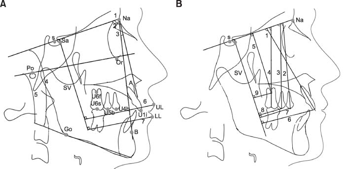
Fig. 2 A, Cephalometric landmarks and skeletal measurements. Landmarks: S, sella; Sa, intersection of the anterior wall of sella turcica and the anterior clinoid processes; Na, nasion; Or, orbitale; Po, porion; A, A point; B, B point; Go, gonion; UL, most anterior point of the upper lip; LL, most anterior point of the lower lip; u6f, furcation point of the upper first molar; u6s, sulcus between buccal tubercules of the upper first molar; u5b, tip of the buccal cusp of the upper second premolar; u4b, tip of the buccal cusp of the upper first premolar; U1i, incisal edge of the upper central incisor; L1i, incisal edge of the lower central incisor. SV indicates vertical reference line. Skeletal measurements: 1, SNA; 2, SNB; 3, ANB; 4, Go-Gn SN; 5, FMA; 6, UL ┴ SV; 7, LL ┴ SV. B, dental angular measurements (°): 1, U1-SN; 2, U4-SN; 3, U5-SN; 4, U6-SN. Dental linear Measurements (mm): 5, U6 ┴ SN; 6, U1 ┴ SV; 7, U4 ┴ SV; 8, U5 ┴ SV; 9, U6 ┴ SV.

Fig. 3 Landmarks, angular and linear measurements on the 3D models. Landmarks: In, tip of the incisive papilla; Bc, tip of the buccal cusp of the upper first molar; Mpc, tip of the mesiopalatal cusp of the upper first molar; Dpc, tip of the distopalatal cusp of the upper first molar. Angular measurements (°): α (rotation of upper right first molar) and β (rotation of upper left first molar). Linear measurement (mm): 1, Intermolar distance.
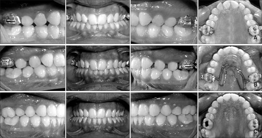
Fig. 4 A, Intraoral photographs before distalization; B, intraoral photographs after distalization; C, posttreatment intraoral photographs.

Fig. 5 A, Lateral cephalometric radiograph before distalization; B, lateral cephalometric radiograph after distalization; C, lateral cephalometric radiograph at the end of treatment.
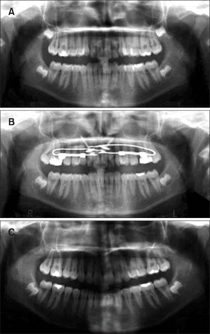
Fig. 6 A, Panoramic radiograph before distalization, B, panoramic radiograph after distalization; C, panoramic radiograph at the end of treatment.

Fig. 7 Superimposition of cephalometric tracing on the maxillary plane. Solid line represents before distalization; broken line represents after distalization.

Fig. 8 Superimposition of 3D image on palatal rugae. White color represents before distalization; gray color represents after distalization.
Cited by 2 articles
-
A reliable method for evaluating upper molar distalization: Superimposition of three-dimensional digital models
Ruhi Nalcaci, Ayse Burcu Kocoglu-Altan, Ali Altug Bicakci, Firat Ozturk, Hasan Babacan
Korean J Orthod. 2015;45(2):82-88. doi: 10.4041/kjod.2015.45.2.82.Three-dimensional analysis of the distal movement of maxillary 1st molars in patients fitted with mini-implant-aided trans-palatal arches
Amirfarhang Miresmaeili, Ahmad Sajedi, Abbas Moghimbeigi, Nasrin Farhadian
Korean J Orthod. 2015;45(5):236-244. doi: 10.4041/kjod.2015.45.5.236.
Reference
-
1. Kircelli BH, Pektaş ZO, Kircelli C. Maxillary molar distalization with a bone-anchored pendulum appliance. Angle Orthod. 2006. 76:650–659.2. Papadopoulos MA. Orthodontic treatment of Class II malocclusion with miniscrew implants. Am J Orthod Dentofac Orthop. 2008. 134:604.e1–604.e16.3. Gulati S, Kharbanda OP, Parkash H. Dental and skeletal changes after intraoral molar distalization with sectional jig assembly. Am J Orthod Dentofacial Orthop. 1998. 114:319–327.
Article4. Gelgör IE, Büyükyilmaz T, Karaman AI, Dolanmaz D, Kalayci A. Intraosseous screw-supported upper molar distalization. Angle Orthod. 2004. 74:838–850.5. Angelieri F, Almeida RR, Almeida MR, Fuziy A. Dentoalveolar and skeletal changes associated with the pendulum appliance followed by fixed orthodontic treatment. Am J Orthod Dentofacial Orthop. 2006. 129:520–527.
Article6. Keles A, Sayınsu K. A new approach in maxillary molar distalization: intraoral bodily molar distalizer. Am J Orthod Dentofacial Orthop. 2000. 117:39–48.
Article7. Keles A, Erverdi N, Sezen S. Bodily distalization of molars with absolute anchorage. Angle Orthod. 2003. 73:471–482.8. Bolla E, Muratore F, Carano A, Bowman SJ. Evaluation of maxillary molar distalization with the distal jet: a comparison with other contemporary methods. Angle Orthod. 2002. 72:481–494.9. Escobar SA, Tellez PA, Moncada CA, Villegas CA, Latorre CM, Oberti G. Distalization of maxillary molars with the bone-supported pendulum: a clinical study. Am J Orthod Dentofacial Orthop. 2007. 131:545–549.
Article10. Chiu PP, McNamara JA Jr, Franchi L. A comparison of two intraoral molar distalization appliances: distal jet versus pendulum. Am J Orthod Dentofacial Orthop. 2005. 128:353–365.
Article11. Doruk C, Ağar U, Babacan H. The role of the headgear timer in extraoral co-operation. Eur J Orthod. 2004. 26:289–291.
Article12. Ağar U, Doruk C, Biçakçi AA, Büküsoğlu N. The role of psycho-social factors in headgear compliance. Eur J Orthod. 2005. 27:263–267.
Article13. Locatelli R, Bednar J, Dietz VS, Gianelly AA. Molar distalization with superelastic NiTi wire. J Clin Orthod. 1992. 26:277–279.14. Wilson WL, Wilson RC. Multi-directional 3D functional Class II treatment. J Clin Orthod. 1987. 21:186–189.15. Hilgers JJ. The pendulum appliance for Class II non-compliance therapy. J Clin Orthod. 1992. 26:706–714.16. Ghosh J, Nanda RS. Evaluation of an intraoral maxillary molar distalization technique. Am J Orthod Dentofacial Orthop. 1996. 110:639–646.
Article17. Byloff FK, Darendeliler MA. Distal molar movement using the pendulum appliance. Part 1: Clinical and radiological evaluation. Angle Orthod. 1997. 67:249–260.18. Kinzinger GS, Fritz UB, Sander FG, Diedrich PR. Efficiency of a pendulum appliance for molar distalization related to second and third molar eruption stage. Am J Orthod Dentofacial Orthop. 2004. 125:8–23.
Article19. Jones RD, White JM. Rapid Class II molar correction with an open coil jig. J Clin Orthod. 1992. 26:661–664.20. Runge ME, Martin JT, Bukai F. Analysis of rapid maxillary molar distal movement without patient cooperation. Am J Orthod Dentofacial Orthop. 1999. 115:153–157.
Article21. Carano A, Testa M. The distal jet for upper molar distalization. J Clin Orthod. 1996. 30:374–380.22. Ngantung V, Nanda RS, Bowman SJ. Posttreatment evaluation of the distal jet appliance. Am J Orthod Dentofacial Orthop. 2001. 120:178–185.
Article23. Keles A. Maxillary unilateral molar distalization with sliding mechanics: a preliminary investigation. Eur J Orthod. 2001. 23:507–515.
Article24. Keles A, Pamukcu B, Tokmak EC. Bilateral maxillary molar distalization with sliding mechanics: Keles slider. World J Orthod. 2002. 3:57–66.25. Björk A, Skieller V. Normal and abnormal growth of the mandible. A synthesis of longitudinal cephalometric implant studies over a period of 25 years. Eur J Orthod. 1983. 5:1–46.
Article26. Dahlberg G. Statistical methods for medical and biological students. 1948. London: Allen and Unwin, Ltd;9–232.27. Turley PK, Kean C, Schur J, Stefanac J, Gray J, Hennes J, Poon LC. Orthodontic force application to titanium endosseous implants. Angle Orthod. 1988. 58:151–162.28. Roberts WE, Helm FR, Marshall KJ, Gongloff RK. Rigid endosseous implants for orthodontic and orthopedic anchorage. Angle Orthod. 1989. 59:247–256.29. Linder-Aronson S, Nordenram A, Anneroth G. Titanium implant anchorage in orthodontic treatment: an experimental investigation in monkeys. Eur J Orthod. 1990. 12:414–419.
Article30. Smalley WM, Shapiro PA, Hohl TH, Kokich VG, Branemark PI. Osseointegrated titanium implants for maxillofacial protraction in monkeys. Am J Orthod Dentofacial Orthop. 1988. 94:285–295.
Article31. Kinzinger GS, Gülden N, Yildizhan F, Diedrich PR. Efficiency of a skeletonized distal jet appliance supported by miniscrew anchorage for noncompliance maxillary molar distalization. Am J Orthod Dentofacial Orthop. 2009. 136:578–586.
Article32. Bondemark L, Kurol J, Bernhold M. Repelling magnets versus superelastic nickel-titanium coils in the simultaneous distal movement of maxillary first and second molars. Angle Orthod. 1994. 64:189–198.33. Worms FW, Isaacson RJ, Speidel TM. A concept and classifications of centers of rotation and extraoral force systems. Angle Orthod. 1973. 43:384–401.34. Gianelly AA, Vaitas AS, Thomas WM. The use of magnets to move molars distally. Am J Orthod Dentofacial Orthop. 1989. 96:161–167.
Article35. Gianelly AA. Distal movement of the maxillary molars. Am J Orthod Dentofacial Orthop. 1998. 114:66–72.
Article36. Ten Hoeve A. Palatal bar and lip bumper in nonextraction treatment. J Clin Orthod. 1985. 19:272–291.37. Jeckel N, Rakosi T. Molar distalization by intra-oral force application. Eur J Orthod. 1991. 3:43–46.
Article38. Smith DI. The eruption of third molars following extraction of second molars. Dental Pract. 1958. 8:292–294.39. Staggers JA. A comparison of results of second molar and first premolar extraction treatment. Am J Orthod Dentofacial Orthop. 1990. 98:430–436.
Article
- Full Text Links
-
- Actions
-
Cited
- CITED
-
- Close
- Share
-
- Similar articles
-
- Immediate changes in the mandibular dentition after maxillary molar distalization using headgear
- Zygoma-gear appliance for intraoral upper molar distalization
- Evaluation of the effects of the third molar on distalization and the effects of attachments on distalization and expansion with clear aligners: Three-dimensional finite element study
- Anatomical factors of the maxillary tuberosity that influence molar distalization
- Combined treatment with headgear and the Frog appliance for maxillary molar distalization: a randomized controlled trial
