Korean J Orthod.
2024 Jul;54(4):239-246. 10.4041/kjod24.017.
Anatomical factors of the maxillary tuberosity that influence molar distalization
- Affiliations
-
- 1Department of Orthodontics, Universidad del Valle, Cali, Colombia
- 2Clínica Imbanaco, Research Center, Cali, Colombia
- KMID: 2558293
- DOI: http://doi.org/10.4041/kjod24.017
Abstract
Objective
To examine the areas of the maxillary tuberosity (MT) (coronal, apical, width, and height) with respect to the presence or absence of the third molar to establish possible anatomical limitations for molar distalization.
Methods
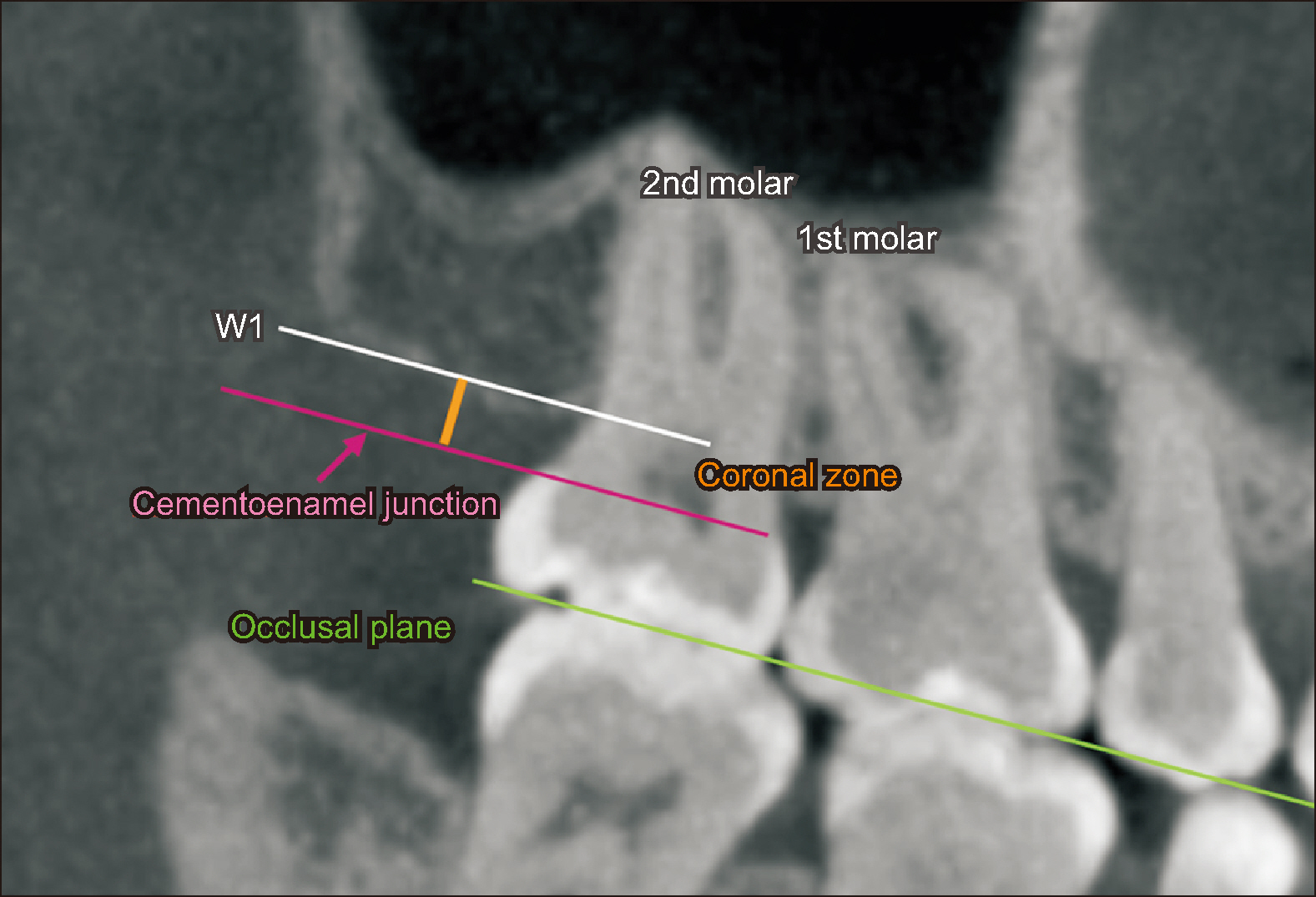
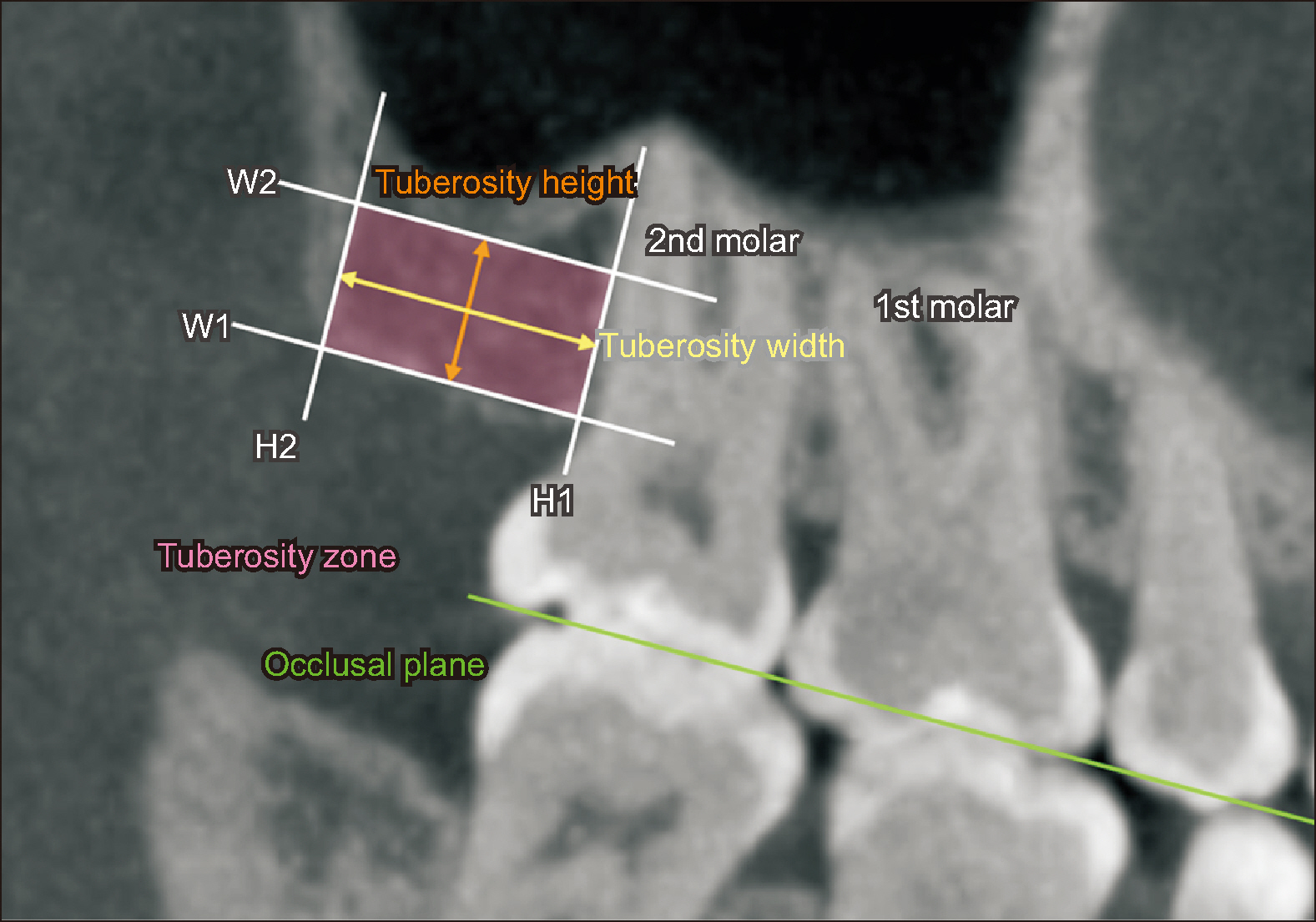
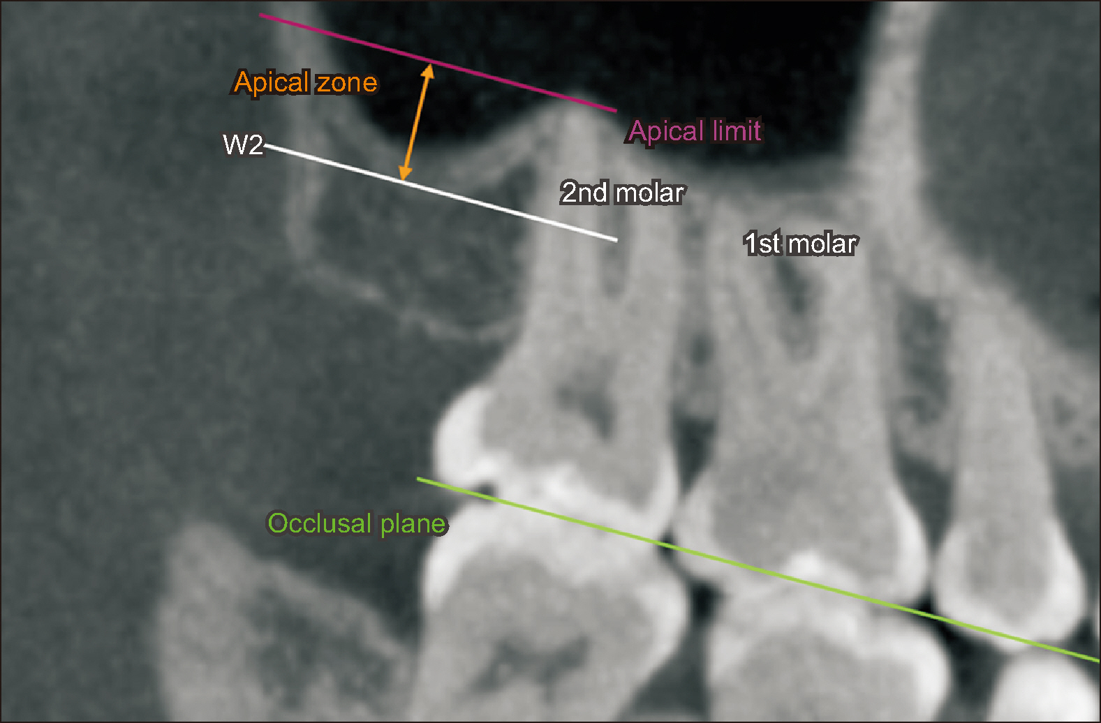
A total of 277 tuberosities were evaluated through sagittal computed tomography (CT) images, divided for measurement into coronal (free of bone), apical (area of influence of the maxillary sinus), and tuberosity (bony area) zones, and stratified by the presence or absence of the third molar, sex, and two age subgroups. Mann–Whitney U test was used to compare the groups considering the third molar.
Results
The medians of the width and height of the tuberosity decreased significantly in the absence of the third molar (P < 0.001). The apical area also showed differences, with negative values in the absence of the third molar and positive values in the presence of the third molar (P < 0.001). However, no differences were observed for the coronal area (P > 0.05).
Conclusions
In the absence of the third molar, the size of the MT, represented by its width and height, was smaller and negative values (decrease) were observed for the maxillary sinus. The sagittal CT provides useful information regarding the amount of bone tissue available for distalization and relationship of the second molar with respect to the maxillary sinus, which allows individualizing each case in relation to the amount and type of movement expected.
Figure
Reference
-
References
1. Cheung LK, Fung SC, Li T, Samman N. 1998; Posterior maxillary anatomy: implications for Le Fort I osteotomy. Int J Oral Maxillofac Surg. 27:346–51. https://doi.org/10.1016/s0901-5027(98)80062-3. DOI: 10.1016/S0901-5027(98)80062-3. PMID: 9804196.
Article2. Apinhasmit W, Chompoopong S, Methathrathip D, Sangvichien S, Karuwanarint S. 2005; Clinical anatomy of the posterior maxilla pertaining to Le Fort I osteotomy in Thais. Clin Anat. 18:323–9. https://doi.org/10.1002/ca.20131. DOI: 10.1002/ca.20131. PMID: 15971227.
Article3. Bayome M, Park JH, Bay C, Kook YA. 2021; Distalization of maxillary molars using temporary skeletal anchorage devices: a systematic review and meta-analysis. Orthod Craniofac Res. 24 Suppl 1:103–12. https://doi.org/10.1111/ocr.12470. DOI: 10.1111/ocr.12470. PMID: 33484608.
Article4. Chae JM. 2006; A new protocol of Tweed-Merrifield directional force technology with microimplant anchorage. Am J Orthod Dentofacial Orthop. 130:100–9. https://doi.org/10.1016/j.ajodo.2005.10.020. DOI: 10.1016/j.ajodo.2005.10.020. PMID: 16849080.5. Garib DG, Yatabe MS, Ozawa TO, Silva Filho OG. 2010; Alveolar bone morphology under the perspective of the computed tomography: defining the biological limits of tooth movement. Dental Press J Orthod. 15:192–205. Português. https://pesquisa.bvsalud.org/portal/resource/pt/lil-562911. DOI: 10.1590/S2176-94512010000500023. PMID: d2fe53efb95a45d2b5a8ab6eef782798.6. Rosa WGN, de Almeida-Pedrin RR, Oltramari PVP, de Castro Conti ACF, Poleti TMFF, Shroff B, et al. 2023; Total arch maxillary distalization using infrazygomatic crest miniscrews in the treatment of Class II malocclusion: a prospective study. Angle Orthod. 93:41–8. https://doi.org/10.2319/050122-326.1. DOI: 10.2319/050122-326.1. PMID: 36126679. PMCID: PMC9797147.
Article7. Ravera S, Castroflorio T, Garino F, Daher S, Cugliari G, Deregibus A. 2016; Maxillary molar distalization with aligners in adult patients: a multicenter retrospective study. Prog Orthod. 17:12. https://doi.org/10.1186/s40510-016-0126-0. DOI: 10.1186/s40510-016-0126-0. PMID: 27041551. PMCID: PMC4834290. PMID: 970deba192ee417f921a2b9ccfcdac13.
Article8. Marcelino V, Baptista S, Marcelino S, Paço M, Rocha D, Gonçalves MDP, et al. 2023; Occlusal changes with clear aligners and the case complexity influence: a longitudinal cohort clinical study. J Clin Med. 12:3435. https://doi.org/10.3390/jcm12103435. DOI: 10.3390/jcm12103435. PMID: 37240538. PMCID: PMC10219537. PMID: 55db70ba4aa74e718498c3f48149cd3b.9. Karatas OH, Toy E. 2014; Three-dimensional imaging techniques: a literature review. Eur J Dent. 8:132–40. https://doi.org/10.4103/1305-7456.126269. DOI: 10.4103/1305-7456.126269. PMID: 24966761. PMCID: PMC4054026.10. Manojna NL, Sunil G, Ramya K, Ranganayakulu I, Raghu Ram R. 2023; Three-dimensional assessment and comparison of the maxillary tuberosity between skeletal and dental Class I and Class II adults in maxillary third molar agenesis using cone beam computed tomography: a descriptive cross-sectional human study. Cureus. 15:e42232. https://doi.org/10.7759/cureus.42232. DOI: 10.7759/cureus.42232. PMID: 37605685. PMCID: PMC10440149.
Article11. Hui VLZ, Xie Y, Zhang K, Chen H, Han W, Tian Y, et al. 2022; Anatomical limitations and factors influencing molar distalization. Angle Orthod. 92:598–605. https://doi.org/10.2319/092921-731.1. DOI: 10.2319/092921-731.1. PMID: 35604682. PMCID: PMC9374358.12. Chiu PP, McNamara JA Jr, Franchi L. 2005; A comparison of two intraoral molar distalization appliances: distal jet versus pendulum. Am J Orthod Dentofacial Orthop. 128:353–65. https://doi.org/10.1016/j.ajodo.2004.04.031. DOI: 10.1016/j.ajodo.2004.04.031. PMID: 16168332.
Article13. Abdelhady NA, Tawfik MA, Hammad SM. 2020; Maxillary molar distalization in treatment of angle class II malocclusion growing patients: uncontrolled clinical trial. Int Orthod. 18:96–104. https://doi.org/10.1016/j.ortho.2019.11.003. DOI: 10.1016/j.ortho.2019.11.003. PMID: 31974060.
Article14. Ishida T, Yoon HS, Ono T. 2013; Asymmetrical distalization of maxillary molars with zygomatic anchorage, improved superelastic nickel-titanium alloy wires, and open-coil springs. Am J Orthod Dentofacial Orthop. 144:583–93. https://doi.org/10.1016/j.ajodo.2012.10.028. DOI: 10.1016/j.ajodo.2012.10.028. PMID: 24075667.
Article15. Raghis TR, Alsulaiman TMA, Mahmoud G, Youssef M. 2022; Efficiency of maxillary total arch distalization using temporary anchorage devices (TADs) for treatment of Class II-malocclusions: a systematic review and meta-analysis. Int Orthod. 20:100666. https://doi.org/10.1016/j.ortho.2022.100666. DOI: 10.1016/j.ortho.2022.100666. PMID: 35871982.
Article16. Mao B, Tian Y, Xiao Y, Li J, Zhou Y. 2023; The effect of maxillary molar distalization with clear aligner: a 4D finite-element study with staging simulation. Prog Orthod. 24:16. https://doi.org/10.1186/s40510-023-00468-1. DOI: 10.1186/s40510-023-00468-1. PMID: 37183221. PMCID: PMC10183381. PMID: 21ecd418be8b4de78db3c527f063f796.17. Spena R, Turatti G. 2011; Upper molar distalization and periodontally facilitated orthodontics. Rev Española Ortod. 41:246–54. Spanish. https://www.revistadeortodoncia.com/frame_esp.php?id=1149.18. Manzanera E, Llorca P, Manzanera D, García-Sanz V, Sada V, Paredes-Gallardo V. 2018; Anatomical study of the maxillary tuberosity using cone beam computed tomography. Oral Radiol. 34:56–65. https://doi.org/10.1007/s11282-017-0284-x. DOI: 10.1007/s11282-017-0284-x. PMID: 30484092.
Article19. Gapski R, Satheesh K, Cobb CM. 2006; Histomorphometric analysis of bone density in the maxillary tuberosity of cadavers: a pilot study. J Periodontol. 77:1085–90. https://doi.org/10.1902/jop.2006.050118. DOI: 10.1902/jop.2006.050118. PMID: 16734586.
Article20. Pei J, Liu J, Chen Y, Liu Y, Liao X, Pan J. 2020; Relationship between maxillary posterior molar roots and the maxillary sinus floor: cone-beam computed tomography analysis of a western Chinese population. J Int Med Res. 48:300060520926896. https://doi.org/10.1177/0300060520926896. DOI: 10.1177/0300060520926896. PMID: 32489120. PMCID: PMC7278324. PMID: 8d1d327d98c84a958ab6e6d636b077e0.
Article21. Aarts BE, Convens J, Bronkhorst EM, Kuijpers-Jagtman AM, Fudalej PS. 2015; Cessation of facial growth in subjects with short, average, and long facial types- implications for the timing of implant placement. J Craniomaxillofac Surg. 43:2106–11. https://doi.org/10.1016/j.jcms.2015.10.013. DOI: 10.1016/j.jcms.2015.10.013. PMID: 26548528.
Article22. Zachrisson BU. 2005; Bjorn U. Zachrisson, DDS, MSD, PhD, on current trends in adult treatment, part 2. Interview by Robert G. Keim. J Clin Orthod. 39:285–96. quiz 315https://pubmed.ncbi.nlm.nih.gov/15961889/.23. Park JH, Tai K, Kanao A, Takagi M. 2014; Space closure in the maxillary posterior area through the maxillary sinus. Am J Orthod Dentofacial Orthop. 145:95–102. https://doi.org/10.1016/j.ajodo.2012.07.020. DOI: 10.1016/j.ajodo.2012.07.020. PMID: 24373659.24. Oh H, Herchold K, Hannon S, Heetland K, Ashraf G, Nguyen V, et al. 2014; Orthodontic tooth movement through the maxillary sinus in an adult with multiple missing teeth. Am J Orthod Dentofacial Orthop. 146:493–505. https://doi.org/10.1016/j.ajodo.2014.03.025. DOI: 10.1016/j.ajodo.2014.03.025. PMID: 25263152.25. Kim S, Lee NK, Park JH, Ku JH, Kim Y, Kook YA, et al. 2022; Treatment effects after maxillary total arch distalization using a modified C-palatal plate in patients with Class II malocclusion with sinus pneumatization. Am J Orthod Dentofacial Orthop. 162:469–76. https://doi.org/10.1016/j.ajodo.2021.04.033. DOI: 10.1016/j.ajodo.2021.04.033. PMID: 35773112.
Article26. Sun W, Xia K, Huang X, Cen X, Liu Q, Liu J. 2018; Knowledge of orthodontic tooth movement through the maxillary sinus: a systematic review. BMC Oral Health. 18:91. https://doi.org/10.1186/s12903-018-0551-1. DOI: 10.1186/s12903-018-0551-1. PMID: 29792184. PMCID: PMC5966888. PMID: 932a8c9a278843fea3e84b62bbc486e0.
Article
- Full Text Links
- Actions
-
Cited
- CITED
-
- Close
- Share
- Similar articles
-
- Immediate changes in the mandibular dentition after maxillary molar distalization using headgear
- Zygoma-gear appliance for intraoral upper molar distalization
- Combined treatment with headgear and the Frog appliance for maxillary molar distalization: a randomized controlled trial
- Evaluation of the effects of the third molar on distalization and the effects of attachments on distalization and expansion with clear aligners: Three-dimensional finite element study
- The frog appliance for upper molar distalization: a case report


