A torque-measuring micromotor provides operator independent measurements marking four different density areas in maxillae
- Affiliations
-
- 1Department of Dentistry, Vita e Salute San Raffaele University, Milano, Italy and Private Practitioner, Milan Italy.
- 2Private Practitioner, Vimercate, Italy.
- 3Department of Medical, Oral and Biotechnological Sciences, University of Chieti-Pescara, Chieti, Italy. v.perrotti@unich.it
- KMID: 1974891
- DOI: http://doi.org/10.4047/jap.2015.7.1.51
Abstract
- PURPOSE
Bone density at implant placement site is a key factor to obtain the primary stability of the fixture, which, in turn, is a prognostic factor for osseointegration and long-term success of an implant supported rehabilitation. Recently, an implant motor with a bone density measurement probe has been introduced. The aim of the present study was to test the objectiveness of the bone densities registered by the implant motor regardless of the operator performing them.
MATERIALS AND METHODS
A total of 3704 bone density measurements, performed by means of the implant motor, were registered by 39 operators at different implant sites during routine activity. Bone density measurements were grouped according to their distribution across the jaws. Specifically, four different areas were distinguished: a pre-antral (between teeth from first right maxillary premolar to first left maxillary premolar) and a sub-antral (more distally) zone in the maxilla, and an interforaminal (between and including teeth from first left mandibular premolar to first right mandibular premolar) and a retroforaminal (more distally) zone in the lower one. A statistical comparison was performed to check the inter-operators variability of the collected data.
RESULTS
The device produced consistent and operator-independent bone density values at each tooth position, showing a reliable bone-density measurement.
CONCLUSION
The implant motor demonstrated to be a helpful tool to properly plan implant placement and loading irrespective of the operator using it.
MeSH Terms
Figure
-

Fig. 1 The bone density measurement probe.
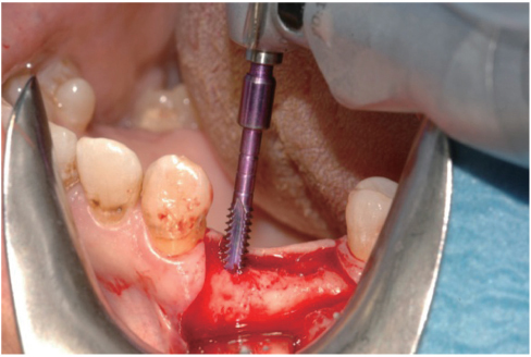
Fig. 2 At measurement, the probe deepens into a 2.3 mm diameter tunnel at the placement site.

Fig. 3 The torque/depth plot generated by the device. I = integral (corresponding to the area below the curve); CM = average torque; N = progressive measurement number; P = depth; CP = maximum torque. GO/STOP = pre-set depth the probe stops at.
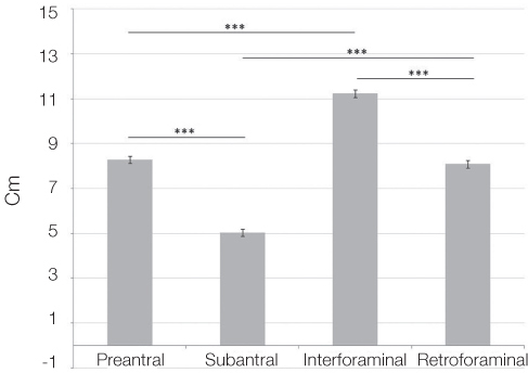
Fig. 4 Statistical comparison of overall means of bone density measurement collected in the four anatomical zones, a pre-antral (anterior) and a sub-antral (posterior) zone in the upper maxilla, and an interforaminal (anterior) and a retroforaminal (posterior) zone in the lower jaw. *: statistically significant difference Cm: average torque
Cited by 1 articles
-
The effect of undersizing and tapping on bone to implant contact and implant primary stability: A histomorphometric study on bovine ribs
Danilo Alessio Di Stefano, Vittoria Perrotti, Gian Battista Greco, Claudia Cappucci, Paolo Arosio, Adriano Piattelli, Giovanna Iezzi
J Adv Prosthodont. 2018;10(3):227-235. doi: 10.4047/jap.2018.10.3.227.
Reference
-
1. Davies JE. Mechanisms of endosseous integration. Int J Prosthodont. 1998; 11:391–401.2. Romanos GE. Bone quality and the immediate loading of implants-critical aspects based on literature, research, and clinical experience. Implant Dent. 2009; 18:203–209.3. Bahat O, Sullivan RM. Parameters for successful implant integration revisited part I: immediate loading considered in light of the original prerequisites for osseointegration. Clin Implant Dent Relat Res. 2010; 12:e2–e12.4. Turkyilmaz I, Ozan O, Yilmaz B, Ersoy AE. Determination of bone quality of 372 implant recipient sites using Hounsfield unit from computerized tomography: a clinical study. Clin Implant Dent Relat Res. 2008; 10:238–244.5. Shapurian T, Damoulis PD, Reiser GM, Griffin TJ, Rand WM. Quantitative evaluation of bone density using the Hounsfield index. Int J Oral Maxillofac Implants. 2006; 21:290–297.6. Turkyilmaz I, Tözüm TF, Tumer C. Bone density assessments of oral implant sites using computerized tomography. J Oral Rehabil. 2007; 34:267–272.7. Valiyaparambil JV, Yamany I, Ortiz D, Shafer DM, Pendrys D, Freilich M, Mallya SM. Bone quality evaluation: comparison of cone beam computed tomography and subjective surgical assessment. Int J Oral Maxillofac Implants. 2012; 27:1271–1277.8. Mah P, Reeves TE, McDavid WD. Deriving Hounsfield units using grey levels in cone beam computed tomography. Dentomaxillofac Radiol. 2010; 39:323–335.9. Nomura Y, Watanabe H, Honda E, Kurabayashi T. Reliability of voxel values from cone-beam computed tomography for dental use in evaluating bone mineral density. Clin Oral Implants Res. 2010; 21:558–562.10. Lekholm U, Zarb GA. Patient selection and preparation. In : Branemark PI, Zarb GA, Albrektsson T, editors. Tissue-integrated prostheses: osseointegration in clinical dentistry. Chicago: Quintessence;1985. p. 199–209.11. Misch CE. Density of Bone: effects on surgical approach and healing. In : Misch CE, editor. Contemporary Implant Dentistry. St. Louis: CV Mosby;2008. p. 645–667.12. Trisi P, Rao W. Bone classification: clinical-histomorphometric comparison. Clin Oral Implants Res. 1999; 10:1–7.13. Iezzi G, Scarano A, Di Stefano D, Arosio P, Doi K, Ricci L, Piattelli A, Perrotti V. Correlation between the bone density recorded by a computerized implant motor and by a histomorphometric analysis: a preliminary in vitro study on bovine ribs. Clin Implant Dent Relat Res. 2015; 17:e35–e44.14. Di Stefano DA, Arosio P, Pagnutti S. A possible novel objective intraoperative measurement of maxillary bone density. Minerva Stomatol. 2013; 62:259–265.15. Ekfeldt A, Christiansson U, Eriksson T, Lindén U, Lundqvist S, Rundcrantz T, Johansson LA, Nilner K, Billström C. A retrospective analysis of factors associated with multiple implant failures in maxillae. Clin Oral Implants Res. 2001; 12:462–467.16. Javed F, Romanos GE. The role of primary stability for successful immediate loading of dental implants. A literature review. J Dent. 2010; 38:612–620.
- Full Text Links
-
- Actions
-
Cited
- CITED
-
- Close
- Share
-
- Similar articles
-
- Screw Insertional Torque Measurement in Spine Surgery: Correlation With Bone Mineral Density and Hounsfield Unit
- A study on accuracy and application of the implant torque controller used in dental clinic
- A Biomechanical Study of Two Kinds of Tapered Pedicle Screws in Osteoporotic Lumber Spine
- The effect of undersizing and tapping on bone to implant contact and implant primary stability: A histomorphometric study on bovine ribs
- Bone density relationship of mandible and cervical vertebrae in panoramic radiography
