The effect of undersizing and tapping on bone to implant contact and implant primary stability: A histomorphometric study on bovine ribs
- Affiliations
-
- 1Dental School, Vita-Salute University IRCCS San Raffaele, Milan, Italy.
- 2Department of Medical, Oral and Biotechnological Sciences, Chieti-Pescara University “G. D'Annunzioâ€, Chieti, Italy. v.perrotti@unich.it
- 3Private Practitioner, Trezzano Sul Naviglio, Milano, Italy.
- 4Private Practitioner, Vimercate, Monza-Brianza, Italy.
- KMID: 2413492
- DOI: http://doi.org/10.4047/jap.2018.10.3.227
Abstract
- PURPOSE
Implant site preparation may be adjusted to achieve the maximum possible primary stability. The aim of this investigation was to study the relation among bone-to-implant contact at insertion, bone density, and implant primary stability intra-operatively measured by a torque-measuring implant motor, when implant sites were undersized or tapped.
MATERIALS AND METHODS
Undersized (n=14), standard (n=13), and tapped (n=13) implant sites were prepared on 9 segments of bovine ribs. After measuring bone density using the implant motor, 40 implants were placed, and their primary stability assessed by measuring the integral of the torque-depth insertion curve. Bovine ribs were then processed histologically, the bone-to-implant contact measured and statistically correlated to bone density and the integral.
RESULTS
Bone-to-implant contact and the integral of the torque-depth curve were significantly greater for undersized sites than tapped sites. Moreover, a correlation between bone to implant contact, the integral and bone density was found under all preparation conditions. The slope of the bone-to-implant/density and integral/density lines was significantly greater for undersized sites, while those corresponding to standard prepared and tapped sites did not differ significantly.
CONCLUSION
The integral of the torque-depth curve provided reliable information about bone-to-implant contact and primary implant stability even in tapped or undersized sites. The linear relations found among the parameters suggests a connection between extent and modality of undersizing and the corresponding increase of the integral and, consequently, of primary stability. These results might help the physician determine the extent of undersizing needed to achieve the proper implant primary stability, according to the planned loading protocol.
Keyword
MeSH Terms
Figure
-
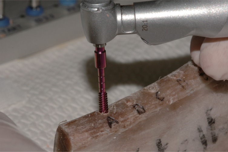
Fig. 1 The probe used to measure the bone density of the bone specimen. The probe has an inverse cone shape, allowing the torque measurement needed to win the friction exerted on the bone wall by its first thread. The average of all torque instantaneous torque values measured throughout its descent, Cm, has been showed to be a reliable measurement of bone density.
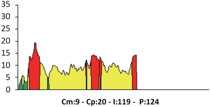
Fig. 2 An example of torque-depth plot showed by the implant motor both when it measures bone density and at implant insertion. Cm, average torque. Cp, peak torque. I, integral of the torque-depth curve. P, depth reached by the probe/implant.
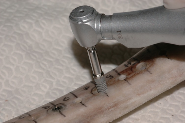
Fig. 3 Implant positioning. Implant placement was performed without irrigation.
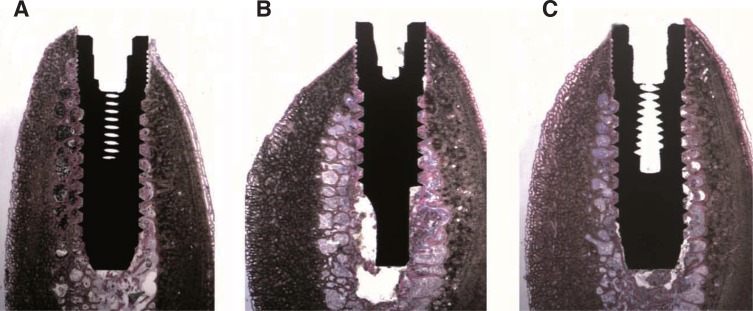
Fig. 4 Low power, histological image of the retrieved specimens belonging to (A) Underprepared site: bone tissue and bone chips surrounding almost the whole implant surface could be observed; only in the portions where the spongy bone was present, and thin bone trabeculae in close proximity with the implant surface were evident; (B) Normal preparation: small bone trabeculae surrounding the implant surface could be seen; (C) Tapped site: small bone trabeculae and bone chips surrounding the implant surface could be seen. (under toluidine blue and acid fuchsin. Original magnification ×6)
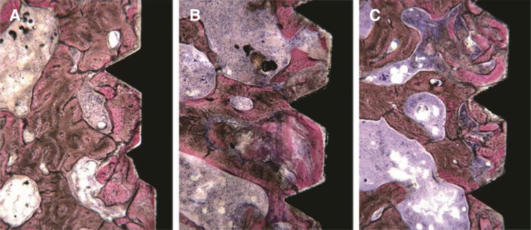
Fig. 5 High power, histological image of the retrieved specimens belonging to (A) Underprepared site: bone tissue in contact with the implant surface could be observed. The bone structure seemed intact; only in some fields, small bone chips were present. (B) Normal preparation: the implant was in contact with small bone trabeculae, which were not damaged in some fields, whilst in other they appeared slightly deformed and followed the implant concavities. (C) Tapped site: many bone chips close to the implant surface could be observed. Only in some fields, bone trabeculae in contact with the implant could be seen. (Under toluidine blue and acid fuchsin. Original magnification ×40)
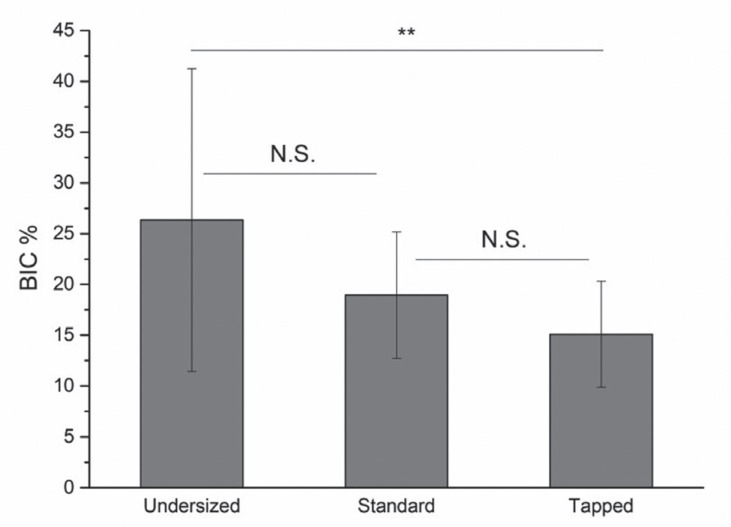
Fig. 6 Average BIC of implants placed into undersized, standard-prepared, and tapped sites. The average BIC corresponding to undersized sites is significantly greater than that corresponding to tapped ones, but is not significantly different from that corresponding to standard-prepared sites. Also the average BIC corresponding to standard-prepared sites is not significantly different from that of tapped sites.

Fig. 7 Average Integral (I) measured at placement of implants in undersized, standard-prepared, and tapped sites. The average (I) corresponding to undersized sites is significantly greater than that corresponding to both standard-prepared and tapped ones. The average (I) corresponding to standard-prepared sites is significantly greater than that corresponding to tapped sites.

Fig. 8 Plot showing the correlation between the BIC and the bone density at the sites of implant placement. Best fitting lines are also shown. Legend: squares and full line, undersized sites; circles and dash-dot line, standard-prepared sites; triangles and dot line, tapped sites. The slope of the line corresponding to the undersized sites is significantly greater than those of the other two lines, which don't differ significantly between each other.
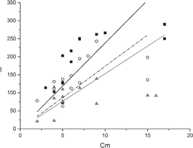
Fig. 9 Plot showing the correlation between the Integral (I) measured at implant placement and the bone density at the sites of implant placement. Best fitting lines are also shown. Legend: squares and full line, undersized sites; circles and dash-dot line, standard-prepared sites. triangles and dot line, tapped sites. The slope of the line corresponding to the undersized sites is significantly greater than those of the other two lines, which don't differ significantly between each other.
Reference
-
1. Glauser R, Sennerby L, Meredith N, Rée A, Lundgren A, Gottlow J, Hämmerle CH. Resonance frequency analysis of implants subjected to immediate or early functional occlusal loading. Successful vs. failing implants. Clin Oral Implants Res. 2004; 15:428–434. PMID: 15248877.
Article2. Möhlhenrich SC, Kniha K, Heussen N, Hölzle F, Modabber A. Effects on primary stability of three different techniques for implant site preparation in synthetic bone models of different densities. Br J Oral Maxillofac Surg. 2016; 54:980–986. PMID: 27461557.
Article3. Friberg B, Ekestubbe A, Mellström D, Sennerby L. Brånemark implants and osteoporosis: a clinical exploratory study. Clin Implant Dent Relat Res. 2001; 3:50–56. PMID: 11441543.
Article4. Stocchero M, Toia M, Cecchinato D, Becktor JP, Coelho PG, Jimbo R. Biomechanical, biologic, and clinical outcomes of undersized implant surgical preparation: A systematic review. Int J Oral Maxillofac Implants. 2016; 31:1247–1263. PMID: 27861649.
Article5. Tabassum A, Walboomers XF, Wolke JG, Meijer GJ, Jansen JA. Bone particles and the undersized surgical technique. J Dent Res. 2010; 89:581–586. PMID: 20212102.
Article6. Shalabi MM, Wolke JG, de Ruijter AJ, Jansen JA. Histological evaluation of oral implants inserted with different surgical techniques into the trabecular bone of goats. Clin Oral Implants Res. 2007; 18:489–495. PMID: 17517059.
Article7. Tabassum A, Meijer GJ, Wolke JG, Jansen JA. Influence of the surgical technique and surface roughness on the primary stability of an implant in artificial bone with a density equivalent to maxillary bone: a laboratory study. Clin Oral Implants Res. 2009; 20:327–332. PMID: 19298286.
Article8. Al-Marshood MM, Junker R, Al-Rasheed A, Al Farraj Aldosari A, Jansen JA, Anil S. Study of the osseointegration of dental implants placed with an adapted surgical technique. Clin Oral Implants Res. 2011; 22:753–759. PMID: 21198894.
Article9. Alghamdi H, Anand PS, Anil S. Undersized implant site preparation to enhance primary implant stability in poor bone density: a prospective clinical study. J Oral Maxillofac Surg. 2011; 69:e506–e512. PMID: 22117707.
Article10. Di Stefano DA, Arosio P, Piattelli A, Perrotti V, Iezzi G. A torque-measuring micromotor provides operator independent measurements marking four different density areas in maxillae. J Adv Prosthodont. 2015; 7:51–55. PMID: 25722838.
Article11. Iezzi G, Scarano A, Di Stefano DA, Arosio P, Doi K, Ricci L, Piattelli A, Perrotti V. Correlation between the bone density recorded by a computerized implant motor and by a histomorphometric analysis: a preliminary in vitro study on bovine ribs. Clin Implant Dent Relat Res. 2015; 17:e35–e44. PMID: 23879771.
Article12. Di Stefano DA, Arosio P. Correlation between bone density and instantaneous torque at implant site preparation: A validation on polyurethane foam blocks of a device assessing density of jawbones. Int J Oral Maxillofac Implants. 2016; 31:e128–e135. PMID: 27632279.
Article13. Degidi M, Daprile G, Piattelli A, Iezzi G. Development of a new implant primary stability parameter: insertion torque revisited. Clin Implant Dent Relat Res. 2013; 15:637–644. PMID: 22008885.
Article14. Iezzi G, Filippone A, Di Stefano DA, Arosio P, Piattelli A, Scarano A, Perrotti V. A site-specific intraoperative measurement of bone-to-implant contact during implant insertion: A study on bovine ribs using a computerized implant motor. J Dent Sci. 2015; 10:21–27.
Article15. Capparé P, Vinci R, Di Stefano DA, Traini T, Pantaleo G, Gherlone EF, Gastaldi G. Correlation between initial BIC and the insertion torque/depth integral recorded with an instantaneous torque-measuring implant motor: An in vivo study. Clin Implant Dent Relat Res. 2015; 17:e613–e620. PMID: 25876078.
Article16. Di Stefano DA, Arosio P, Gastaldi G, Gherlone E. The insertion torque-depth curve integral as a measure of implant primary stability: An in vitro study on polyurethane foam blocks. J Prosthet Dent. 2017; 7. 08.17. Wang TM, Lee MS, Wang JS, Lin LD. The effect of implant design and bone quality on insertion torque, resonance frequency analysis, and insertion energy during implant placement in low or low- to medium-density bone. Int J Prosthodont. 2015; 28:40–47. PMID: 25588172.
Article18. Degidi M, Daprile G, Piattelli A. Influence of underpreparation on primary stability of implants inserted in poor quality bone sites: an in vitro study. J Oral Maxillofac Surg. 2015; 73:1084–1088. PMID: 25861691.19. Piattelli A, Scarano A, Quaranta M. High-precision, cost-effective cutting system for producing thin sections of oral tissues containing dental implants. Biomaterials. 1997; 18:577–579. PMID: 9105598.
Article20. Andrade JM, Estévez-Pérez MG. Statistical comparison of the slopes of two regression lines: A tutorial. Anal Chim Acta. 2014; 838:1–12. PMID: 25064237.
Article21. Ku HH. Notes on the use of propagation of error formulas. J Res Natl Bur Stand. 1966; 70C:263–273.
Article22. Fernandes Ede L, Unikowski IL, Teixeira ER, da Costa NP, Shinkai RS. Primary stability of turned and acid-etched screw-type implants: a removal torque and histomorphometric study in rabbits. Int J Oral Maxillofac Implants. 2007; 22:886–892. PMID: 18271369.
- Full Text Links
-
- Actions
-
Cited
- CITED
-
- Close
- Share
-
- Similar articles
-
- Effect of bone quality and implant surgical technique on implant stability quotient (ISQ) value
- The effect of osteotome technique on primary implant stability according to implant fixture diameter
- The effect of implant shape and bone preparation on primary stability
- Influence of implant fixture design on implant primary stability
- A study on the finite element analysis and bone formation of the apically expandable implant
