Synovial Osteochondromatosis of the Cervical Spine: A Case Report
- Affiliations
-
- 1Department of Radiology, Seoul National University Bundang Hospital, Seongnam, Korea. joonwoo2@gmail.com
- 2Department of Orthopedic Surgery, Seoul National University Bundang Hospital, Seongnam, Korea.
- 3Department of Pathology, Seoul National University Bundang Hospital, Seongnam, Korea.
- KMID: 1941786
- DOI: http://doi.org/10.3348/jksr.2014.70.5.379
Abstract
- Synovial osteochondromatosis is a rare, benign condition characterized by formation of cartilaginous nodules within the synovium. It rarely occurs at cervical spine, and only six cases have been previously reported in the English literature. We describe another case of synovial osteochondromatosis in the cervical spine in a 77-year-old man who presented with compressive myelopathy. Here we briefly review the literature and discuss the differential diagnosis based on CT and MR findings.
MeSH Terms
Figure
-
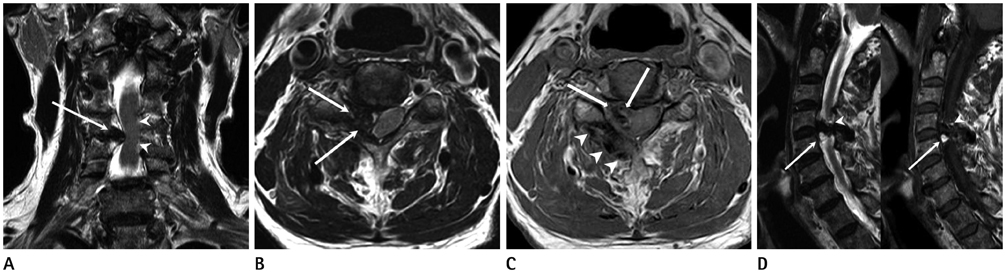
Fig. 1 Preoperative findings of a cervical MRI in a 77-year-old male patient. A. Coronal sectional image of T2-weighted cervical MR shows exophyting mass indenting the dural sac (white arrow) with focal short segmental high signal intensity of the spinal cord (white arrowheads) at the C4/5 level, suggestive of compressive myelopathy. B. Axial sectional image of T2-weighted cervical MR shows low signal intensity of the exophyting mass in the right epidural space at the C4/5 facet joint (white arrows). C. Axial sectional image of T1-weighted cervical MR also shows low to intermediate signal intensity of the exophyting mass in the right epidural space at the C4/5 facet joint (white arrows) and three small sized low signal intensity nodules of similar size posterior to the right lamina of C4 (white arrowheads). D. Sagittal sectional image of both T2 and T1-weighted cervical MR shows focal high signal intensity within the mass, suggesting focal fatty marrow changes (white arrow, white arrowhead).
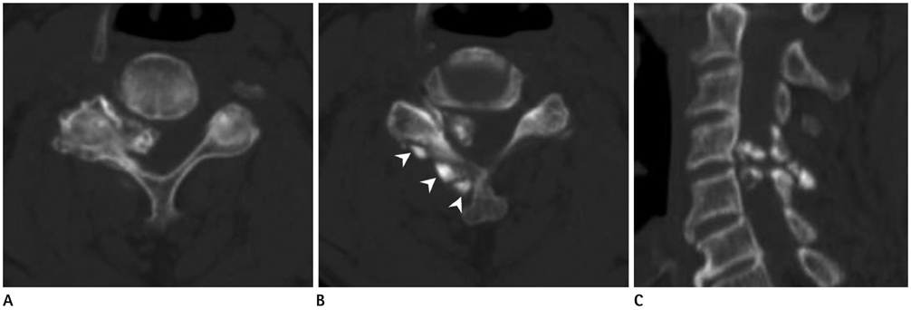
Fig. 2 Preoperative findings of a cervical CT in a 77-year-old male patient. A. Axial sectional image of the preoperative CT shows a large calcified exophyting mass at the C4/5 facet joint. There are multiple osteophytes and degenerative changes at the right C4/5 facet joint with relatively normal configuration of the left side. B. Three small calcified nodules of similar size were seen at the posterior to the right lamina of C4 in the axial image of the preoperative CT (arrowheads). C. Sagittal sectional image of the preoperative CT shows two large calcified masses in the right epidural at the C4/5 level and small calcified nodules posterior to the right lamina.
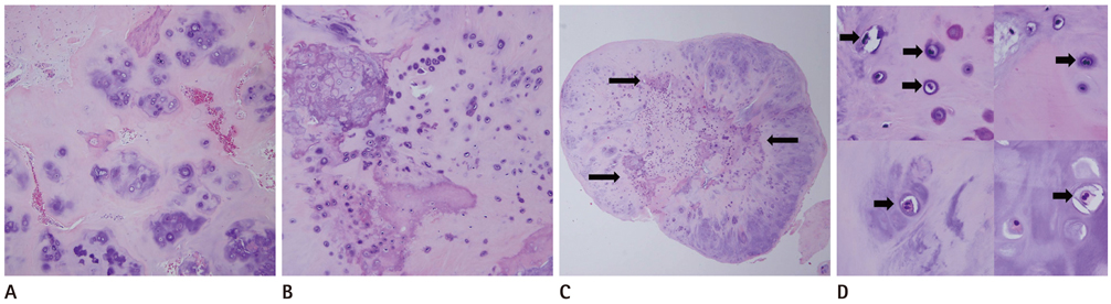
Fig. 3 Histopathologic findings of the cervical mass lesions. A. Note nodules of disorganized cellular metaplastic cartilage in synovial tissue (H&E, × 100). B. Note the disorganized cellular pattern with increased cellularity, cellular atypia and clustering. Neither orderly maturation pattern nor concentric rings of calcification, characteristic in secondary synovial chondromatoses, was noted. C. Note a loose body showing irregular patchy pattern of calcification (arrows) (H&E, × 40). D. Note the frequent binucleated chondrocytes (arrows).
Reference
-
1. Murphey MD, Vidal JA, Fanburg-Smith JC, Gajewski DA. Imaging of synovial chondromatosis with radiologic-pathologic correlation. Radiographics. 2007; 27:1465–1488.2. Fandburg-Smith J. Cartilage and bone forming tumors and tumor-like lesions. In : Miettinen M, editor. Diagnostic Soft Tissue Pathology. New York: Churchill Livingstone;2003. p. 405–411.3. Villacin AB, Brigham LN, Bullough PG. Primary and secondary synovial chondrometaplasia: histopathologic and clinicoradiologic differences. Hum Pathol. 1979; 10:439–451.4. Moody P, Bui MM, Vrionis F, Setzer M, Rojiani AM. Synovial chondromatosis of spine: case report and review of the literature. Ann Clin Lab Sci. 2010; 40:71–74.5. Kyriakos M, Totty WG, Riew KD. Synovial chondromatosis in a facet joint of a cervical vertebra. Spine (Phila Pa 1976). 2000; 25:635–640.6. Greenlee JD, Ghodsi A, Baumbach GL, VanGilder JC. Synovial chondromatosis of the cervical spine. Case illustration. J Neurosurg. 2002; 97:1 Suppl. 150.7. Gallia GL, Weiss N, Campbell JN, McCarthy EF, Tufaro AP, Gokaslan ZL. Vertebral synovial chondromatosis. Report of two cases and review of the literature. J Neurosurg Spine. 2004; 1:211–218.8. Chiba S, Koge N, Oda M, Yamauchi R, Imai T, Matsumoto H, et al. Synovial chondromatosis presenting with cervical radiculopathy: a case report. Spine (Phila Pa 1976). 2003; 28:E396–E400.9. Dharmadhikari R, Dildey P, Hide IG. A rare cause of spinal cord compression: imaging appearances of gout of the cervical spine. Skeletal Radiol. 2006; 35:942–945.10. Feydy A, Lioté F, Carlier R, Chevrot A, Drapé JL. Cervical spine and crystal-associated diseases: imaging findings. Eur Radiol. 2006; 16:459–468.11. Kramer J, Recht M, Deely DM, Schweitzer M, Pathria MN, Gentili A, et al. MR appearance of idiopathic synovial osteochondromatosis. J Comput Assist Tomogr. 1993; 17:772–776.12. Motamedi K, Murphey MD, Fetsch JF, Furlong MA, Vinh TN, Laskin WB, et al. Villonodular synovitis (PVNS) of the spine. Skeletal Radiol. 2005; 34:185–195.
- Full Text Links
-
- Actions
-
Cited
- CITED
-
- Close
- Share
-
- Similar articles
-
- Primary Synovial Osteochondromatosis: A case report
- Synovial Osteochondromatosis of the Subtalar Joint in an Adolescent Baseball Player
- Brown-Sequard Syndrome Caused by a Cervical Synovial Cyst
- A Case of Cervical Synovial Cyst Causing Myelopathy
- Regrowing Synovial Chondromatosis in a Cervical Facet Joint with Radiculopathy
