Computed Tomography Findings of Alveolar Adenoma of the Lung with Histopathologic Comparison: A Case Report
- Affiliations
-
- 1Department of Radiology, Gangneung Asan Hospital, College of Medicine, University of Ulsan, Gangneung, Korea. ryu@gnah.co.kr
- 2Department of Pathology, Gangneung Asan Hospital, College of Medicine, University of Ulsan, Gangneung, Korea.
- 3Department of Thoracic and Cardiovascular Surgery, Gangneung Asan Hospital, College of Medicine, University of Ulsan, Gangneung, Korea.
- KMID: 1941779
- DOI: http://doi.org/10.3348/jksr.2014.70.5.347
Abstract
- Alveolar adenoma is a rare pulmonary neoplasm with a female predominance, and it was considered to be a histologic variant of sclerosing hemangioma in the past. A chest X-ray usually shows a well-defined, peripheral, solitary nodule similar to that of sclerosing hemangioma. Chest CT shows a solitary, well-defined, peripheral nodule with homogeneous density and no contrast enhancement, which is contrary to marked contrast enhancement of sclerosing hemangioma. We report the first case of alveolar adenoma with spotty enhancement of the nodule similar to that of sclerosing hemangioma on contrast enhanced CT, based on the presence of stromal vessels in the interstitium of the compact alveolar area on histopathologic comparison.
MeSH Terms
Figure
-
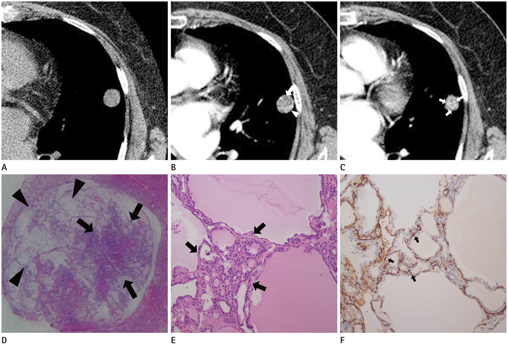
Fig. 1 A 57-year-old woman with alveolar adenoma. A. HRCT with mediastinal setting shows lung nodule with homogenous and low-attenuation (15 Hounsfield unit, HU) compared with that of chest wall muscle (25 HU). The lung nodule with smooth margin is located in the subpleural portion of the lingular segment of the left upper lobe, measuring 1.5 cm in size. B. Contrast enhanced CT shows heterogenous attenuation with mainly low attenuation (25 HU) in the center portion (arrow) and focal enhancement (67 HU) in the left lower lateral portion (arrowhead) of the nodule. C. Contrast enhanced CT shows spotty enhancement (70 HU) in the lower portion of the nodule (arrows). D. Photomicrograph (H&E stain; magnification: × 1) shows a round nodule consisting of stromal vessels of the solid area (arrows) in left half of nodule and cystic component (arrowheads) in right half of nodule. E. Dilated cystic alveolar area alternating with compacted small alveolar area (arrows). The compact small alveolar area has higher density of stromal vessels than that of cystic area (H&E, × 100). The final pathologic report was alveolar adenoma. F. CD34 immunohistochemical stain (× 100) highlighting the difference of stromal vessel density between dilated cystic alveolar area and compact small alveolar area. Stromal vessel is stained as dark brown channel or line (arrows).
Reference
-
1. Yousem SA, Hochholzer L. Alveolar adenoma. Hum Pathol. 1986; 17:1066–1071.2. Semeraro D, Gibbs AR. Pulmonary adenoma: a variant of sclerosing haemangioma of lung? J Clin Pathol. 1989; 42:1222–1223.3. Fujimoto K, Müller NL, Sadohara J, Harada H, Hayashi A, Hayabuchi N. Alveolar adenoma of the lung: computed tomography and magnetic resonance imaging findings. J Thorac Imaging. 2002; 17:163–166.4. Nosotti M, Mendogni P, Rosso L, Tosi D, Palleschi A, Basciu M, et al. Alveolar adenoma of the lung: unusal diagnosis of a lesion positive on PET scan. A case report. J Cardiothorac Surg. 2012; 7:1–4.5. Sak SD, Koseoglu RD, Demirag F, Akbulut H, Gungor A. Alveolar adenoma of the lung. Immunohistochemical and flow cytometric characteristics of two new cases and a review of the literature. APMIS. 2007; 115:1443–1449.6. Kim GY, Kim J, Choi YS, Kim HJ, Ahn G, Han J. Sixteen cases of sclerosing hemangioma of the lung including unusual presentations. J Korean Med Sci. 2004; 19:352–358.7. Burke LM, Rush WI, Khoor A, Mackay B, Oliveira P, Whitsett JA, et al. Alveolar adenoma: a histochemical, immunohistochemical, and ultrastructural analysis of 17 cases. Hum Pathol. 1999; 30:158–167.8. Halldorsson A, Dissanaike S, Kaye KS. Alveolar adenoma of the lung: a clinicopathological description of a case of this very unusual tumour. J Clin Pathol. 2005; 58:1211–1214.9. Petrella F, Rizzo S, Pelosi G, Borri A, Galetta D, Gasparri R, et al. Giant alveolar adenoma causing severe dyspnoea. J Thorac Oncol. 2010; 5:1088–1090.10. Chung MJ, Lee KS, Han J, Sung YM, Chong S, Kwon OJ. Pulmonary sclerosing hemangioma presenting as solitary pulmonary nodule: dynamic CT findings and histopathologic comparisons. AJR Am J Roentgenol. 2006; 187:430–437.
- Full Text Links
-
- Actions
-
Cited
- CITED
-
- Close
- Share
-
- Similar articles
-
- Ground-Glass Opacity in Lung Metastasis from Adenocarcinoma of the Stomach: A Case Report
- A Case of Alveolar Adenoma Involving Multiple Lung Nodules
- Pulmonary Metastases of Alveolar Soft-Part Sarcoma: CT Findings in Three Patients
- Rapid Progression of Metastatic Pulmonary Calcification and Alveolar Hemorrhage in a Patient with Chronic Renal Failure and Primary Hyperparathyroidism
- A Case of Pulmonary Alveolar Proteinosis with Spontaneous Resolution
