Rapid Progression of Metastatic Pulmonary Calcification and Alveolar Hemorrhage in a Patient with Chronic Renal Failure and Primary Hyperparathyroidism
- Affiliations
-
- 1Department of Radiology, Chosun University College of Medicine, Gwangju, Korea. dhk0827@chosun.ac.kr
- 2Department of Internal Medicine, Chosun University College of Medicine, Gwangju, Korea.
- 3Department of Anaesthesiology and Pain Medicine, Seonam University College of Medicine, Namwon, Korea.
- KMID: 2002898
- DOI: http://doi.org/10.3348/jksr.2013.68.6.473
Abstract
- Metastatic pulmonary calcification (MPC) is common in patients with chronic renal failure. The authors experienced a patient with chronic renal failure and primary hyperparathyroidism by parathyroid adenoma accompanied with rapid progressions of MPC and alveolar hemorrhage. Recent chest radiographs, compared with previous chest radiographs, showed rapid accumulation of calcification in both upper lungs. Following up on the high-resolution CT scan after five years demonstrates more increased nodules in size and ground glass opacity. The patient was diagnosed with MPC and alveolar hemorrhage by transbronchial lung biopsy. We assumed rapid progression of MPC and alveolar hemorrhage in underlying chronic renal failures could be a primary hyperparathyroidism which may be caused by parathyroid adenoma detected incidentally. Therefore parathyroid adenoma was treated with ethanol injections. Herein, we have reported on CT findings of MPC with alveolar hemorrhage and reviewed our case along with other articles.
MeSH Terms
Figure
-
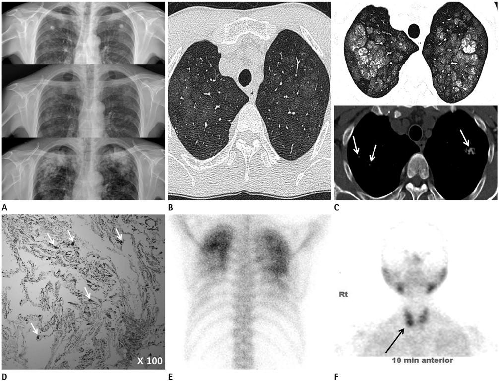
Fig. 1 A 50-year-old male with chronic renal failure having metastatic pulmonary calcification. A. Chest radiographs (upper image five years ago, middle and lower images during recent six months, respectively) show more prominent multiple nodules with calcification and rapid progression of metastatic pulmonary calcification in both mid- and upper-lung fields during the recent several months. B. HRCT five years ago shows ill-defined nodules with ground glass opacity in both upper lung fields without calcification. C. HRCT scan (upper image) at last admission shows poorly defined nodular opacities and ground glass opacities. Mediastinal window image with no contrast injection (lower image) demonstrate definite nodular calcifications (arrows) compared with the previous CT scan. D. H&E stained section of the lesion obtained by bronchoscopic biopsy shows calcium deposits as black spots in the alveolar wall and bronchiolar wall (arrows). E. 99mTcO4 bone scan shows localized radiotracer accumulation representing metastatic pulmonary calcification in both upper lung fields. F. 99mTc-MIBI parathyroid scan shows localized hyperdense accumulation demonstrating adenoma at the right lower pole of the thyroid (arrow). Note.-HRCT = high-resolution CT, MIBI = methy-isobutyl-isonitrile
Reference
-
1. Hartman TE, Müller NL, Primack SL, Johkoh T, Takeuchi N, Ikezoe J, et al. Metastatic pulmonary calcification in patients with hypercalcemia: findings on chest radiographs and CT scans. AJR Am J Roentgenol. 1994; 162:799–802.2. Sanders C, Frank MS, Rostand SG, Rutsky EA, Barnes GT, Fraser RG. Metastatic calcification of the heart and lungs in end-stage renal disease: detection and quantification by dual-energy digital chest radiography. AJR Am J Roentgenol. 1987; 149:881–887.3. Romagnoli M, Mourad G, Serre I, Senac JP, Paradis L, Godard Ph, et al. Diffuse pulmonary calcinosis without calcium metabolism abnormalities in a renal transplant recipient. Eur Respir J. 1997; 10:958–960.4. Marchiori E, Müller NL, Souza AS Jr, Escuissato DL, Gasparetto EL, de Cerqueira EM. Unusual manifestations of metastatic pulmonary calcification: high-resolution CT and pathological findings. J Thorac Imaging. 2005; 20:66–70.5. Jost RG, Sagel SS. Metastatic calcification in the lung apex. AJR Am J Roentgenol. 1979; 133:1188–1190.6. Kuhlman JE, Ren H, Hutchins GM, Fishman EK. Fulminant pulmonary calcification complicating renal transplantation: CT demonstration. Radiology. 1989; 173:459–460.7. Franquet T, Lee KS, Müller NL. Thin-section CT findings in 32 immunocompromised patients with cytomegalovirus pneumonia who do not have AIDS. AJR Am J Roentgenol. 2003; 181:1059–1063.8. Collard HR, Schwarz MI. Diffuse alveolar hemorrhage. Clin Chest Med. 2004; 25:583–592. vii
- Full Text Links
-
- Actions
-
Cited
- CITED
-
- Close
- Share
-
- Similar articles
-
- A Case of Primary Hyperparathyroidism Associated with Hypercalcemic Crisis and Systemic Calcinosis
- Metastatic pulmonalry calcification associated with malignant lymphoma: a case report
- A Case of Metastatic Gastric Calcification in Acute Renal Failure
- Metastatic Calcification of the Finger in a Chronic Renal Failure Patient
- A Case of Metastatic Pulmonary Calcification in Primary Hyperparathyroidism
