Extrinsic Tracheal Compression Caused by Scoliosis of the Thoracic Spine and Chest Wall Deformity: A Case Report
- Affiliations
-
- 1Department of Radiology, St. Paul's Hospital, College of Medicine, The Catholic University of Korea, Seoul, Korea. leebae@catholic.ac.kr
- 2Department of Pulmonology, St. Paul's Hospital, College of Medicine, The Catholic University of Korea, Seoul, Korea.
- KMID: 1941778
- DOI: http://doi.org/10.3348/jksr.2014.70.5.343
Abstract
- Extrinsic airway compression due to chest wall deformity is not commonly observed. Although this condition can be diagnosed more easily with the help of multidetector CT, the standard treatment method has not yet been definitely established. We report a case of an eighteen-year-old male who suffered from severe extrinsic tracheal compression due to scoliosis and straightening of the thoracic spine, confirmed on CT and bronchoscopy. The patient underwent successful placement of tracheal stent but later died of bleeding from the tracheostomy site probably due to tracheo-brachiocephalic artery fistula. We describe the CT and bronchoscopic findings of extrinsic airway compression due to chest wall deformity as well as the optimal treatment method, and discuss the possible explanation for bleeding in the patient along with review of the literature.
MeSH Terms
Figure
-
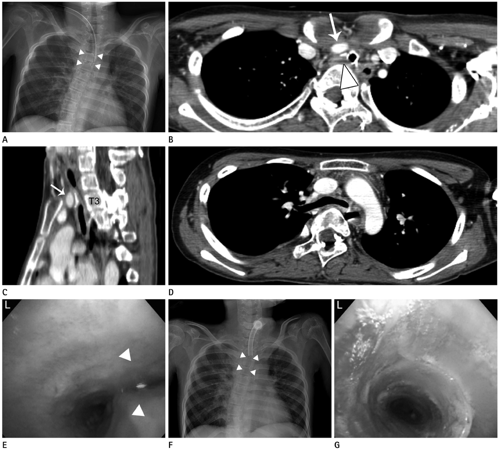
Fig. 1 18-year-old male with intractable respiratory distress. A. Initial chest radiograph at our hospital shows severe scoliosis and nonvisualization of a segment of distal trachea (arrowheads) caudal to the intubation tube. The carina can still be seen. B. Axial contrast-enhanced CT image at the level of T3 vertebra and upper end of sternal manubrium shows markedly narrowed mediastinal space and resultant extrinsic compression of the trachea (arrowhead) especially by the brachiocephalic artery (arrow) that courses between the manubrium and the vertebral body. C. Sagittal reconstructed CT image clearly demonstrates the straightened thoracic spine and extremely narrow space between the sternal manubrium and T3 vertebra. Note how brachiocephalic artery (arrow) also contributes to the compression of the trachea in between. D. Axial contrast-enhanced CT image at the level of T5 vertebra shows splaying of the lower trachea by the vertebral body. E. Bronchoscopy before the tracheal stent placement shows the extrinsic compression of the distal trachea, especially on the right side (arrowheads). F. Chest radiograph after the placement of the silicone stent shows the stent just above the carina, maintaining patency (arrowheads). G. Bronchoscopy performed four days after the stent insertion shows well-placed silicone stent inside the distal trachea. Note.-L = left
Reference
-
1. Donnelly LF, Bisset GS 3rd. Airway compression in children with abnormal thoracic configuration. Radiology. 1998; 206:323–326.2. Karoll M, Hernandez RJ, Wessel HU. Computed tomography diagnosis of bronchial compression by the spine after surgical correction of scoliosis. Pediatr Radiol. 1984; 14:335–336.3. Grillo HC, Wright CD, Dartevelle PG, Wain JC, Murakami S. Tracheal compression caused by straight back syndrome, chest wall deformity, and anterior spinal displacement: techniques for relief. Ann Thorac Surg. 2005; 80:2057–2062.4. Tatekawa Y, Muraji T. Surgical strategy for acquired tracheomalacia due to innominate artery compression of the trachea. Eur J Cardiothorac Surg. 2011; 39:412–413.5. Brigger MT, Boseley ME. Management of tracheal stenosis. Curr Opin Otolaryngol Head Neck Surg. 2012; 20:491–496.6. Keats TE, Sistrom C. Atlas of Radiologic Measurement. 7th ed. St. Louis: Mosby;2001. p. 124–125.7. Tsakiridis K, Darwiche K, Visouli AN, Zarogoulidis P, Machairiotis N, Christofis C, et al. Management of complex benign post-tracheostomy tracheal stenosis with bronchoscopic insertion of silicon tracheal stents, in patients with failed or contraindicated surgical reconstruction of trachea. J Thorac Dis. 2012; 4:Suppl 1. 32–40.8. Maeda K, Ono S, Tazuke Y, Baba K. Long-term outcomes of congenital tracheal stenosis treated by metallic airway stenting. J Pediatr Surg. 2013; 48:293–296.9. Ogawa K, Nitta N, Sonoda A, Takahashi M, Suzuki T, Kitamura S, et al. Tracheo-brachiocephalic artery fistula after tracheostomy associated with thoracic deformity: a case report. J Med Case Rep. 2011; 5:595.10. Tatekawa Y, Tojo T, Kanehiro H, Nakajima Y. Multistage approach for tracheobronchomalacia caused by a chest deformity in the setting of severe scoliosis. Surg Today. 2007; 37:910–914.
- Full Text Links
-
- Actions
-
Cited
- CITED
-
- Close
- Share
-
- Similar articles
-
- Surgical Therapy of Airway Compression with Severe Kyphoscoliosis
- Juvenile Idiopathic Scoliosis
- Right Ventricular Compression Observed in Echocardiography from Pectus Excavatum Deformity
- Left Second Rib Exostosis, Spinal Cord Compression and Left Upper Thoracic Scoliosis: A Rare Triad
- Surgical Management of an Isolated Huge Innominate Artery Aneurysm Causing Tracheal Compression: A Case Report
