J Korean Soc Surg Hand.
2014 Mar;19(1):1-6. 10.12790/jkssh.2014.19.1.1.
The Change of the Ulnar Variance in Accordance with the Wrist Position in Ulnocarpal Impaction Syndrome
- Affiliations
-
- 1Department of Orthopedic Surgery, Hanyang University College of Medicine, Seoul, Korea. leegh@hanyang.ac.kr
- 2Department of Orthopedic Surgery, KEPCO Medical Center, Seoul, Korea.
- KMID: 1896920
- DOI: http://doi.org/10.12790/jkssh.2014.19.1.1
Abstract
- PURPOSE
We evaluated the change of the ulnar variance (UV) as forearm rotation in patients with ulnocarpal impaction syndrome (UIS).
METHODS
Twenty patients who suffered from ulnar side pain of the wrist and had abnormal lesions at ulno-basal side of the lunate in the radiologic examinations were included in this study. Their UVs in six wrist position (neutral, supination, pronation, neutral and grip, supination and grip, pronation and grip) were measured by the method of perpendiculars. UVs and the maximum change of UV in patients with UIS were compared with those of control group statistically.
RESULTS
There were statistically significant differences in UVs of all forearm rotation and grip status. The maximum change of UV was in supination position to pronation and grip status for all cases. The mean maximum change of UV in patients with UIS was 2.03+/-1.03 mm, and that of control group was 1.86+/-0.86 mm. But there was no significant difference between them. The ulnar shortening osteotomy was performed for thirteen UIS patients, and one patient with osteoarthritis at distal radio-ulnar joint was operated with Darrach procedure. Six patients underwent conservative treatment.
CONCLUSION
There were no significant differences in the maximum change of UV as forearm rotation between UIS patients and control group.
Figure
-
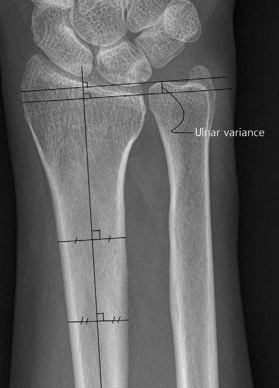
Fig. 1. The method of perpendiculars is used to measure the ulnar variance. The two lines perpendicular to the long axis of the radius are drawn through the distal ulnar aspect of radial volar cortical rim and the dorsal rim of the distal ulna respectively. The distance between these lines is measured as the ulnar variance.
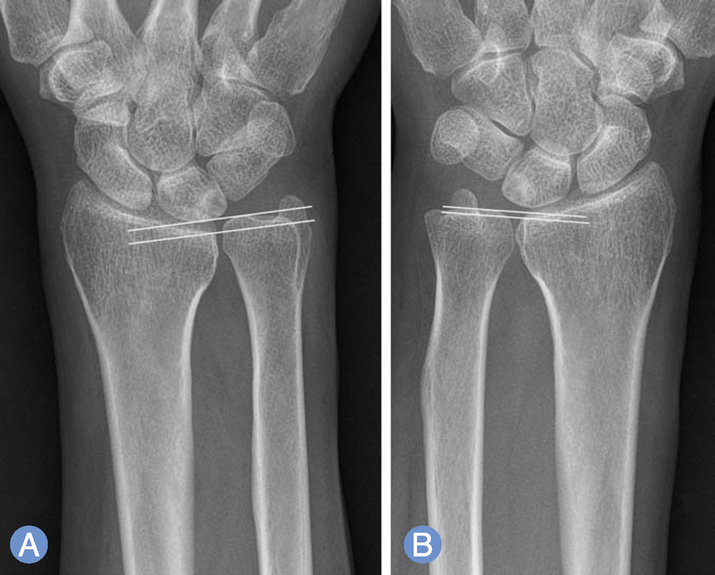
Fig. 2. (A) Maximum ulnar variance is measured when gripping in pronation. (B) Minimum ulnar variance is measured when relaxed in supination.
Reference
-
1. Palmer AK, Werner FW. Biomechanics of the distal radioulnar joint. Clin Orthop Relat Res. 1984; (187):26–35.
Article2. Palmer AK, Werner FW. The triangular fibrocartilage complex of the wrist: anatomy and function. J Hand Surg Am. 1981; 6:153–62.3. Palmer AK, Glisson RR, Werner FW. Relationship between ulnar variance and triangular fibrocartilage complex thickness. J Hand Surg Am. 1984; 9:681–2.
Article4. Jung JM, Baek GH, Kim JH, Lee YH, Chung MS. Changes in ulnar variance in relation to forearm rotation and grip. J Bone Joint Surg Br. 2001; 83:1029–33.
Article5. Tomaino MM. The importance of the pronated grip X-ray view in evaluating ulnar variance. J Hand Surg Am. 2000; 25:352–7.
Article6. Yeh GL, Beredjiklian PK, Katz MA, Steinberg DR, Bozentka DJ. Effects of forearm rotation on the clinical evaluation of ulnar variance. J Hand Surg Am. 2001; 26:1042–6.
Article7. Steyers CM, Blair WF. Measuring ulnar variance: a comparison of techniques. J Hand Surg Am. 1989; 14:607–12.
Article8. Shrout PE, Fleiss JL. Intraclass correlations: uses in assessing rater reliability. Psychol Bull. 1979; 86:420–8.
Article9. Park JC, Sur YJ, Rhee SK, Song SW, Lee SM, Han SH. Ulnar shortening osteotomy for the treatment of ulnar impaction syndrome. J Korean Soc Surg Hand. 2009; 14:172–8.10. Friedman SL, Palmer AK, Short WH, Levinsohn EM, Halperin LS. The change in ulnar variance with grip. J Hand Surg Am. 1993; 18:713–6.
Article11. Makita A, Nakamura T, Takayama S, Toyama Y. The shape of the triangular fibrocartilage during pronation-supination. J Hand Surg Br. 2003; 28:537–45.
Article12. Nishiwaki M, Nakamura T, Nakao Y, Nagura T, Toyama Y. Ulnar shortening effect on distal radioulnar joint stability: a biomechanical study. J Hand Surg Am. 2005; 30:719–26.
Article13. Lee JS, Park MJ. Diagnostic usefulness of ulnocarpal stress for triangular fibrocartilage lesions. J Korean Soc Hand Surg. 2006; 11:1–6.
- Full Text Links
-
- Actions
-
Cited
- CITED
-
- Close
- Share
-
- Similar articles
-
- Ulnar impaction syndrome: how to diagnose and treat?
- Arthroscopy of the Wrist and Ulnar Shortening Osteotomy for the Treatment of the Ulnar Impaction Syndrome
- Updates on Ulnar Impaction Syndrome
- Intraoperative Arthroscopic Findings of Ulnar Impaction Syndrome
- Positive or Negative Ulnar Variance after Ulnar Shortening for Ulnar Impaction Syndrome: A Retrospective Study
