Clin Orthop Surg.
2012 Sep;4(3):216-220. 10.4055/cios.2012.4.3.216.
Positive or Negative Ulnar Variance after Ulnar Shortening for Ulnar Impaction Syndrome: A Retrospective Study
- Affiliations
-
- 1Department of Orthopaedic Surgery, Chungnam National University School of Medicine, Daejeon, Korea. hyunsd@cnu.ac.kr
- KMID: 1392984
- DOI: http://doi.org/10.4055/cios.2012.4.3.216
Abstract
- BACKGROUND
The goal of this study was to compare simple radiographic findings and clinical results according to residual ulnar variance following ulnar shortening for ulnar impaction syndrome.
METHODS
Forty-five cases of ulnar impaction syndrome, which were treated with ulnar shortening from 2005 to 2008, were studied retrospectively. Group I included 13 cases with positive residual variance after ulnar shortening and group II included 32 cases with negative variance after shortening. The presence of a lunate cystic lesion both preoperatively and at final follow-up and assessments of wrist function based on the modified Mayo wrist score, the disabilities of the arm, shoulder, and hand (DASH) score, as well as the Chun and Palmer score were evaluated.
RESULTS
A cystic lesion of the lunate was present in 4 cases preoperatively and the size decreased in 2 cases at final follow-up in group I, and in 10 and 5 cases, respectively, in group II. No statistical difference was observed between the groups. The modified Mayo wrist score, DASH score, as well as the Chun and Palmer score improved significantly in both groups. No significant differences were observed between the two groups in terms of the proportion of positive cystic lesions at final follow-up or the functional scores.
CONCLUSIONS
After ulnar shortening, the degree of radiological change in the cystic lunate lesions and clinical improvement did not differ significantly between the groups with unintended residual positive and negative variance after shortening.
MeSH Terms
Figure
-
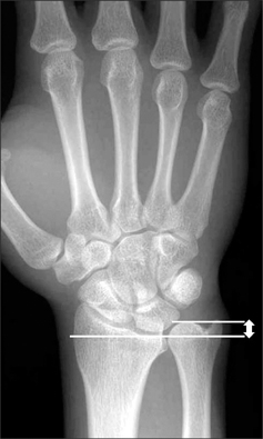
Fig. 1 The ulnar variance was measured by drawing a line through the volar sclerotic line of the distal radius perpendicular to its longitudinal axis. The variance is the distance between this line and the distal cortical rim of the ulnar dome.
Reference
-
1. Chun S, Palmer AK. The ulnar impaction syndrome: follow-up of ulnar shortening osteotomy. J Hand Surg Am. 1993. 18(1):46–53.
Article2. Lauder AJ, Luria S, Trumble TE. Oblique ulnar shortening osteotomy with a new plate and compression system. Tech Hand Up Extrem Surg. 2007. 11(1):66–73.
Article3. Feldon P, Terrono AL, Belsky MR. Wafer distal ulna resection for triangular fibrocartilage tears and/or ulna impaction syndrome. J Hand Surg Am. 1992. 17(4):731–737.
Article4. Wehbe MA, Cautilli DA. Ulnar shortening using the AO small distractor. J Hand Surg Am. 1995. 20(6):959–964.
Article5. Constantine KJ, Tomaino MM, Herndon JH, Sotereanos DG. Comparison of ulnar shortening osteotomy and the wafer resection procedure as treatment for ulnar impaction syndrome. J Hand Surg Am. 2000. 25(1):55–60.
Article6. Loh YC, Van Den Abbeele K, Stanley JK, Trail IA. The results of ulnar shortening for ulnar impaction syndrome. J Hand Surg Br. 1999. 24(3):316–320.
Article7. Palmer AK, Glisson RR, Werner FW. Ulnar variance determination. J Hand Surg Am. 1982. 7(4):376–379.
Article8. Fleiss JL, Cohen J. The equivalence of weighted kappa and the intraclass correlation coefficient as measures of reliability. Educ Psychol Meas. 1973. 33(3):613–619.
Article9. Cooney WP, Bussey R, Dobyns JH, Linscheid RL. Difficult wrist fractures. Perilunate fracture-dislocations of the wrist. Clin Orthop Relat Res. 1987. (214):136–147.10. Hudak PL, Amadio PC, Bombardier C. Development of an upper extremity outcome measure: the DASH (disabilities of the arm, shoulder and hand): the Upper Extremity Collaborative Group (UECG). Am J Ind Med. 1996. 29(6):602–608.
Article11. Friedman SL, Palmer AK. The ulnar impaction syndrome. Hand Clin. 1991. 7(2):295–310.
Article12. Tomaino MM. Ulnar impaction syndrome in the ulnar negative and neutral wrist: diagnosis and pathoanatomy. J Hand Surg Br. 1998. 23(6):754–757.13. Palmer AK. Triangular fibrocartilage complex lesions: a classification. J Hand Surg Am. 1989. 14(4):594–606.
Article14. Palmer AK, Werner FW. Biomechanics of the distal radioulnar joint. Clin Orthop Relat Res. 1984. (187):26–35.
Article15. Tomaino MM, Rubin DA. The value of the pronated-grip view radiograph in assessing dynamic ulnar positive variance: a case report. Am J Orthop (Belle Mead NJ). 1999. 28(3):180–181.16. Escobedo EM, Bergman AG, Hunter JC. MR imaging of ulnar impaction. Skeletal Radiol. 1995. 24(2):85–90.
Article17. Imaeda T, Nakamura R, Shionoya K, Makino N. Ulnar impaction syndrome: MR imaging findings. Radiology. 1996. 201(2):495–500.
Article18. Tatebe M, Nakamura R, Horii E, Nakao E. Results of ulnar shortening osteotomy for ulnocarpal impaction syndrome in wrists with neutral or negative ulnar variance. J Hand Surg Br. 2005. 30(2):129–132.
Article19. Nishiwaki M, Nakamura T, Nakao Y, Nagura T, Toyama Y. Ulnar shortening effect on distal radioulnar joint stability: a biomechanical study. J Hand Surg Am. 2005. 30(4):719–726.
Article
- Full Text Links
-
- Actions
-
Cited
- CITED
-
- Close
- Share
-
- Similar articles
-
- Arthroscopy of the Wrist and Ulnar Shortening Osteotomy for the Treatment of the Ulnar Impaction Syndrome
- Ulnar Shortening Osteotomy for the Treatment of Ulnar Impaction Syndrome
- Updates on Ulnar Impaction Syndrome
- Treatment of Ulnar Impaction Syndrome using Arthroscopy and Ulnar Shortening Osteotomy
- Long-term Outcomes of Ulnar Shortening Osteotomy for Idiopathic Ulnar Impaction Syndrome: At Least 5-Years Follow-up
