Granular Cell Tumor in the Pituitary Stalk: A Case Report
- Affiliations
-
- 1Department of Neurosurgery, Ewha Womans University Mokdong Hospital, Seoul, Korea. drekseo@ewha.ac.kr
- KMID: 1882014
- DOI: http://doi.org/10.14791/btrt.2015.3.1.60
Abstract
- Granular cell tumors (GCTs) have been reported in various tissues, especially the skin and subcutaneous soft tissue of the head and neck. We report a 60-year-old man who presented with intermittent headache and dizziness for 3 months, but no other neurological symptoms. Magnetic resonance imaging (MRI) showed the presence of a mass in the pituitary stalk, and contrast-enhanced MRI showed nodular enhancement in this region. The lesion was completely excised microscopically via a frontotemporal (pterional) approach. On pathological examination, a final diagnosis of a typical GCT was made.
Keyword
MeSH Terms
Figure
-

Fig. 1 Preoperative magnetic resonance imaging findings. A: A preoperative T1-weighted gadolinium-enhanced axial image shows a homogenous enhanced round mass (white arrow). B: A T1-weighted gadolinium-enhanced coronal image shows the pituitary stalk (white arrow). C: An anterior view of image B shows a round mass (white arrowhead). D: A T2-weighted axial image shows a mass (arrow) with low signal intensity.

Fig. 2 Surgical findings. A: The tumor (asterisk) is round, and adjacent to the pituitary stalk. B: After tumor excision, the pituitary stalk (arrow) appears thinned, but remains intact.

Fig. 3 Histopathologic findings. A: The tumor consists of large polygonal cells with ample granular cytoplasm and small, oval, eccentric nuclei (hematoxylin and eosin; original magnification ×100). B: The tumor shows dense fibrocollagenous tissue admixed with granular cell nests. Multifocal lymphocytic infiltration can also be observed (original magnification ×100). C: Immunostaining for S-100 shows diffuse weak to strong positivity (original magnification ×100).
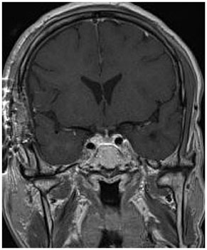
Fig. 4 Postoperative magnetic resonance imaging findings. A postoperative T1-weighted gadolinium-enhanced coronal image shows resection of the tumor adjacent to the pituitary stalk. The pituitary stalk remains intact.
Reference
-
1. Cohen-Gadol AA, Pichelmann MA, Link MJ, et al. Granular cell tumor of the sellar and suprasellar region: clinicopathologic study of 11 cases and literature review. Mayo Clin Proc. 2003; 78:567–573.
Article2. Cone L, Srinivasan M, Romanul FC. Granular cell tumor (choristoma) of the neurohypophysis: two cases and a review of the literature. AJNR Am J Neuroradiol. 1990; 11:403–406.3. Halbauer DJ, Mészáros I, Dóczi T, et al. Rare sellar region tumors. Pathol Oncol Res. 2003; 9:134–137.
Article4. Higuchi M, Tsuji M, Ikeda H. Symptomatic hypophyseal granular cell tumour: endocrinological and clinicopathological analysis. Br J Neurosurg. 1997; 11:582–586.
Article5. Hurley TR, D'Angelo CM, Clasen RA, Wilkinson SB, Passavoy RD. Magnetic resonance imaging and pathological analysis of a pituicytoma: case report. Neurosurgery. 1994; 35:314–317. discussion 317.6. Rhee JS, Wackym PA, Hague K, Wolfe D, King WA. Granular cell tumor of the pituitary fossa. Ann Otol Rhinol Laryngol. 2002; 111:754–758.7. Vogelgesang S, Junge MH, Pahnke J, Gaab MR, Warzok RW. August 2001: Sellar/suprasellar mass in a 59-year-old woman. Brain Pathol. 2002; 12:135–136. 1398. Shizukuishi T, Abe O, Haradome H, Fukushima T, Katayama Y, Sugitani M. Granular cell tumor of the neurohypophysis with optic tract edema. Jpn J Radiol. 2014; 32:179–182.
Article9. Wilkinson MD, Fulham MJ, Besser M. Neuroimaging findings in a suprasellar granular cell tumor. J Comput Assist Tomogr. 2003; 27:26–29.
Article10. Iglesias A, Arias M, Brasa J, Páramo C, Conde C, Fernandez R. MR imaging findings in granular cell tumor of the neurohypophysis: a difficult preoperative diagnosis. Eur Radiol. 2000; 10:1871–1873.
Article11. Takei Y, Seyama S, Pearl GS, Tindall GT. Ultrastructural study of the human neurohypophysis. II. Cellular elements of neural parenchyma, the pituicytes. Cell Tissue Res. 1980; 205:273–287.12. Figarella-Branger D, Dufour H, Fernandez C, Bouvier-Labit C, Grisoli F, Pellissier JF. Pituicytomas, a mis-diagnosed benign tumor of the neurohypophysis: report of three cases. Acta Neuropathol. 2002; 104:313–319.
Article13. Moriyama E, Matsumoto Y, Meguro T, Mano S. Suprasellar granular cell tumor. Neurol Med Chir (Tokyo). 1996; 36:237–240.14. Tomita T, Gates E. Pituitary adenomas and granular cell tumors. Incidence, cell type, and location of tumor in 100 pituitary glands at autopsy. Am J Clin Pathol. 1999; 111:817–825.
Article15. Ellison D, Love S, Chimelli L. Neuropathology: A Reference Text of CNS Pathology. 3rd ed. Philadelphia: Elsevier;2013.16. Nakamura T, Hirato J, Hotchi M, Kyoshima K, Nakamura Y. Astrocytoma with granular cell tumor-like changes. Report of a case with histochemical and ultrastructural characterization of granular cells. Acta Pathol Jpn. 1990; 40:206–211.
Article17. Losa M, Saeger W, Mortini P, et al. Acromegaly associated with a granular cell tumor of the neurohypophysis: a clinical and histological study. Case report. J Neurosurg. 2000; 93:121–126.
Article
- Full Text Links
-
- Actions
-
Cited
- CITED
-
- Close
- Share
-
- Similar articles
-
- Sporadic Hemangioblastoma in the Pituitary Stalk: A Case Report and Review of the Literature
- Pituitary Adenoma Confined Within the Pituitary Stalk: A Case Report and Literature Review
- Surgical Treatment of Hemangioblastoma in the Pituitary Stalk: An Extremely Rare Case
- Atypical Granular Cell Tumor of the Sellar Region
- A Case of Thyroid Granular Cell Tumor
