Development of myenteric plexus in human foetuses: a quantitative study
- Affiliations
-
- 1Department of Anatomy, All India Institute of Medical Sciences, New Delhi, India. seemahkg@gmail.com
- KMID: 1845280
- DOI: http://doi.org/10.5115/acb.2015.48.2.124
Abstract
- Maturation of neurons of the myenteric plexus (MP) of human fetal sigmoid colon was studied at various weeks of gestation (WG). There is abundant literature on the development of MP in various segments of the gut but there are fewer reports on the development of MP in human sigmoid colon which is a site of various disorders. Sigmoid colonic segments from 12 aborted foetuses aged 14-23WG were processed for NADPH histochemistry. Stereologic evaluation of the neuronal cell profiles, numerical density, number of neurons per ganglion and myenteric fraction was conducted using using imageJ software. According to gestational age, foetuses were assigned into two groups (group 1 [n=7], less than <17WG and group 2 [n=5], more than >17WG). The overall size of neuronal cell profiles in the MP was significantly increased (P<0.05). The numerical density of neurons decreased in group 2 in comparison to group 1, the number of neurons per ganglion and myenteric fraction were increased in group 2 but all these were not statistically significant. This study revealed that the maturational event increases after 17WG and extensive innervations is established at 23WG. During prenatal life there is an increase in the neuronal cell size from 14-23WG signifying maturational process. Such studies are essential for clinicians and surgeons to correlate the normal and pathologic development of the enteric nervous system.
MeSH Terms
Figure
-
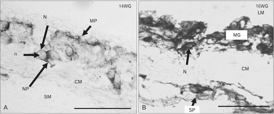
Fig. 1 Microphotograph of cross-section of human foetal sigmoid colon in NADPH-diaphorase stain at 14 weeks of gestation (WG) and 16WG. (A) Myenteric plexus (MP) at 14WG having neurons (N) with round to oval nucleus (n) and scanty cytoplasm. Neuronal processes (NP) can be clearly demarcated. Scattered neurons and thin nerve fibres present in submucosa (SM). (B) There is progressive increase in the sizes of myenteric ganglia (MG) and N at 16WG. Nerve fibres increase in inner circular muscle (CM). There is also gradually increase in neuropil. Submucosal plexus (SP) present in the SM towards the CM. LM, outer longitudinal muscle. Scale bar=50 µm (A, B). Reproduced from J Morphol Sci 2013;30:156-66 [18], with permission of Brazilian Society of Anatomy and Pan-American Association of Anatomy.

Fig. 2 Microphotograph of NADPH-diaphorase stained sigmoid colon at 23 weeks of gestation (WG). (A) At this age, Myenteric ganglia (MG) have increased in size and large number of nerve fibres developed in circular muscle (CM). Submucosal plexus (SP) present in the submucosa (SM) towards the CM. Internodal strand (Is) is seen between two MG. (B) Higher magnification shows large neurons (N) with rounded to oval nuclei (asterix) and extensive neuropil (np) in MG. Thickened neuronal processes (NP) were clearly visible. NF, nerve fibres. Scale bars=200 µm (A), 20 µm (B). Reproduced from J Morphol Sci 2013;30:156-66 [18], with permission of Brazilian Society of Anatomy and Pan-American Association of Anatomy.

Fig. 3 All the parameters (area, perimeter and ferret diameter) which were taken to assess the overall size of the neuronal cell in the myenteric plexus were significantly increased (P=0.02). WG, weeks of gestation; group 1, <17WG; group 2, >17WG. *Statistically significant.
Reference
-
1. Gershon MD. The second brain: the scientific basis of gut instinct and a groundbreaking new understanding of nervous disorders of the stomach and intestines. New York: Harper Collins;1998.2. Furness JB, Costa M. The enteric nervous system. Edinburg: Churchill Livingstone;1987.3. Costa M, Brookes SJ. The enteric nervous system. Am J Gastroenterol. 1994; 89:S129–S137.4. Le Douarin NM. The neural crest. Cambridge: Cambridge University Press;1982.5. Burns AJ, Douarin NM. The sacral neural crest contributes neurons and glia to the post-umbilical gut: spatiotemporal analysis of the development of the enteric nervous system. Development. 1998; 125:4335–4347.6. Serbedzija GN, Burgan S, Fraser SE, Bronner-Fraser M. Vital dye labelling demonstrates a sacral neural crest contribution to the enteric nervous system of chick and mouse embryos. Development. 1991; 111:857–866.7. Kapur RP. Colonization of the murine hindgut by sacral crest-derived neural precursors: experimental support for an evolutionarily conserved model. Dev Biol. 2000; 227:146–155.8. Fekete É, Bagyánszki M, Resch BA. Prenatal development of the myenteric plexus in the human fetal small intestine. Acta Biol Szeged. 2000; 44:3–19.9. Okamoto E, Ueda T. Embryogenesis of intramural ganglia of the gut and its relation to Hirschprung's disease. J Pediatr Surg. 1967; 2:437–443.10. McKeown SJ, Chow CW, Young HM. Development of the submucous plexus in the large intestine of the mouse. Cell Tissue Res. 2001; 303:301–305.11. Wallace AS, Burns AJ. Development of the enteric nervous system, smooth muscle and interstitial cells of Cajal in the human gastrointestinal tract. Cell Tissue Res. 2005; 319:367–382.12. Stach W. Neuronal organization of the myenteric plexus (Auerbach) in the small intestine of the pig. I. Type I neurons. Z Mikrosk Anat Forsch. 1980; 94:833–849.13. Hitchcock RJ, Pemble MJ, Bishop AE, Spitz L, Polak JM. Quantitative study of the development and maturation of human oesophageal innervation. J Anat. 1992; 180(Pt 1):175–183.14. Gershon MD, Erde SM. The nervous system of the gut. Gastroenterology. 1981; 80:1571–1594.15. Mandarim-de-Lacerda CA. Foot length growth related to crown-rump length, gestational age and weight in human staged fresh fetuses. An index for anatomical and medical use. Surg Radiol Anat. 1990; 12:103–107.16. Sailaja K, Ahuja RK, Gopinath G. Biparietal diameter: a useful measure for determining gestational age of human abortuses. Natl Med J India. 1996; 9:165–167.17. Scherer-Singler U, Vincent SR, Kimura H, McGeer EG. Demonstration of a unique population of neurons with NADPH-diaphorase histochemistry. J Neurosci Methods. 1983; 9:229–234.18. Singh S, Shariff A, Roy TS, Kumar H. Prenatal development of the myenteric plexus in human sigmoid colon. J Morphol Sci. 2013; 30:156–166.19. Gabella G. Neuron size and number in the myenteric plexus of the newborn and adult rat. J Anat. 1971; 109:81–95.20. Wallace AS, Barlow AJ, Navaratne L, Delalande JM, Tauszig-Delamasure S, Corset V, Thapar N, Burns AJ. Inhibition of cell death results in hyperganglionosis: implications for enteric nervous system development. Neurogastroenterol Motil. 2009; 21:768–e49.21. Montedonico S, Sri Paran T, Pirker M, Rolle U, Puri P. Developmental changes in submucosal nitrergic neurons in the porcine distal colon. J Pediatr Surg. 2006; 41:1029–1035.
- Full Text Links
-
- Actions
-
Cited
- CITED
-
- Close
- Share
-
- Similar articles
-
- Impact of Myenteric Plexus Alterations on Diabetes Related Gastrointestinal Dysmotility
- Unusual Histology of Eosinophilic Myenteric Ganglionitis: A Case Report
- Immunohistochemical localization of calcium binding proteins and some neurotransmitters in myenteric plexus of goat stomach
- Changes of Immunoreactivities of Calcium Channel alpha(1B)Subunit in Myenteric Plexus of Capsaicin Treated Adult Rats
- Pathogenesis of Achalasia
