Predictive Capability of the Spetzler-Martin versus Supplementary Grading Scale for Microsurgical Outcomes of Cerebellar Arteriovenous Malformations
- Affiliations
-
- 1Department of Neurological Surgery, University of Virginia, Charlottesville, VA, USA. kcl3j@hscmail.mcc.virginia.edu
- KMID: 1810789
- DOI: http://doi.org/10.7461/jcen.2013.15.4.307
Abstract
- The recently described supplementary grading scale may be superior to the widely used Spetzler-Martin grading scale in the prediction of microsurgical outcomes for cerebellar arteriovenous malformations (AVM). We report two cases of ruptured cerebellar AVMs with the same Spetzler-Martin grade but different supplementary grades treated with microsurgical resection. Both patients had symptomatic brainstem compression from cerebellar hematomas and subsequently underwent uncomplicated surgeries; however, their outcomes were significantly different. It has previously been proposed that AVMs distort cerebellar anatomy in a different manner than supratentorial cerebral anatomy thereby potentially resulting in misrepresentation when utilizing the Spetzler-Martin grading scale. However, the components of the supplementary grading scale are independent of cerebellar anatomy, which may explain why it has been shown to be better than the Spetzler-Martin grading scale for prediction of surgical outcomes. In addition, due to the smaller volume of the posterior fossa compared to the supratentorial compartment, rupture of cerebellar AVMs may result in rapid and catastrophic neurological compromise. Therefore, the role of microsurgery may be more critical for AVMs of the cerebellar than for those located elsewhere. Simple and effective grading systems are invaluable tools for clinical and surgical decision-making, although the decisions rendered should always be made in conjunction with the patient's presentation and the physician's experience.
Keyword
MeSH Terms
Figure
-
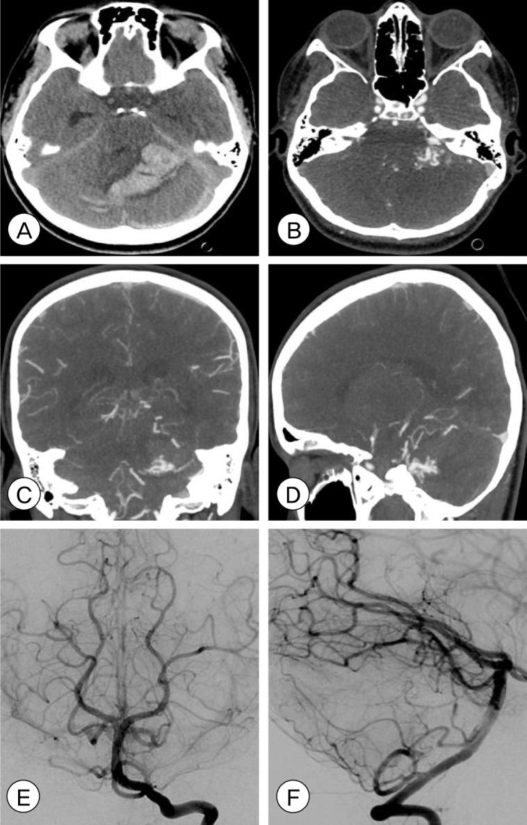
Fig. 1 Pre-operative (A) non-contrast brain computed tomography (CT), axial view, shows a large cerebellar hematoma with severe effacement of the fourth ventricle, resultant hydrocephalus, and tonsillar herniation. Brain CT angiography (CTA), axial (B), coronal (C), and sagittal (D) views shows a Spetzler-Martin Grade II, supplementary grade I left cerebellar arteriovenous malformation (AVM) measuring 12×8 mm in size, supplied by the left superior cerebellar artery (SCA) and anterior inferior cerebellar artery (AICA) and draining into the left transverse and straight sinuses. Post-operative angiography with left vertebral artery injection, AP (E) and lateral (F) views, shows no residual AVM, consistent with complete microsurgical obliteration.
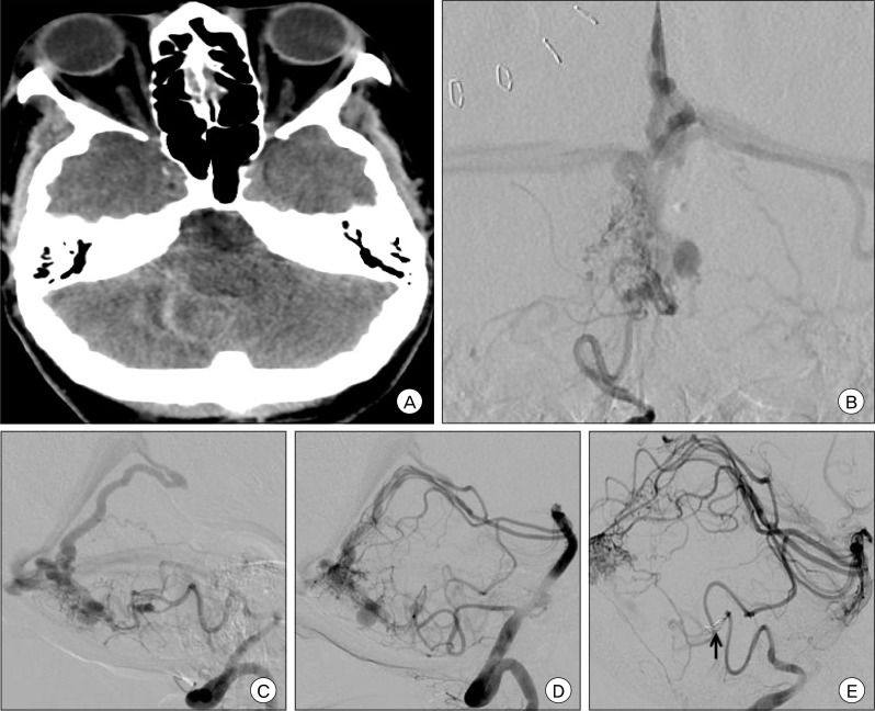
Fig. 2 Pre-operative (A) non-contrast brain CT, axial view, shows diffuse cerebellar subarachnoid hemorrhage and intraventricular hemorrhage of the fourth ventricle resulting in obstructive hydrocephalus. Pre-operative angiography with right vertebral artery injection, AP (B) and lateral (C) views, and left vertebral artery injection, lateral view (D) shows a Spetzler-Martin Grade II, supplementary grade III right cerebellar arteriovenous malformation (AVM) measuring 14×15×13 mm in size, supplied by the bilateral superior cerebellar arteries (SCAs) and posterior inferior cerebellar arteries (PICAs) and draining into the vein of Galen. There are multiple feeding artery aneurysms on the right PICA as well as several venous dilatations of the deep draining vein. After pre-operative polyvinyl alcohol (PVA) particle embolization through the right PICA, the feeding artery was occluded with coils (arrow) as shown on this DSA right vertebral artery injection lateral view (E).
Cited by 3 articles
-
Gamma Knife Radiosurgery for ARUBA-Eligible Patients with Unruptured Brain Arteriovenous Malformations
Byung Sup Kim, Je Young Yeon, Jong-Soo Kim, Seung-Chyul Hong, Hyung Jin Shin, Jung-Il Lee
J Korean Med Sci. 2019;34(36):. doi: 10.3346/jkms.2019.34.e232.Preoperative Embolization of Cerebral Arteriovenous Malformations with Silk Suture and Particles: Technical Considerations and Outcomes
Jordan R. Conger, Dale Ding, Daniel M. Raper, Robert M. Starke, Christopher R. Durst, Kenneth C. Liu, Mary E. Jensen, Avery J. Evans
J Cerebrovasc Endovasc Neurosurg. 2016;18(2):90-99. doi: 10.7461/jcen.2016.18.2.90.Surgical Approaches for Symptomatic Cerebral Cavernous Malformations of the Thalamus and Brainstem
Dale Ding, Robert M. Starke, R. Webster Crowley, Kenneth C. Liu
J Cerebrovasc Endovasc Neurosurg. 2017;19(1):19-35. doi: 10.7461/jcen.2017.19.1.19.
Reference
-
1. Lawton MT, Kim H, McCulloch CE, Mikhak B, Young WL. A supplementary grading scale for selecting patients with brain arteriovenous malformations for surgery. Neurosurgery. 2010; 4. 66(4):702–713. discussion 713. PMID: 20190666.
Article2. Rodríguez-Hernández A, Kim H, Pourmohamad T, Young WL, Lawton MT. University of California, San Francisco Arteriovenous Malformation Study Project. Cerebellar arteriovenous malformations: Anatomic subtypes, surgical results, and increased predictive accuracy of the supplementary grading system. Neurosurgery. 2012; 12. 71(6):1111–1124. PMID: 22986595.
- Full Text Links
-
- Actions
-
Cited
- CITED
-
- Close
- Share
-
- Similar articles
-
- Clinical Analysis of Complications of Surgery for Arteriovenous Malformations of the Brain
- Grading System and Outcome of Surgical Treatment of Arteriovenous Malformations
- Cerebellar Glioblastoma Presenting as a Cerebellar Hemorrhage in a Child
- Feasibility of Gamma Knife Radiosurgery for Brain Arteriovenous Malformations According to Nidus Type
- Surgical Results with Low-Grade Arteriovenous Malformations : A Single Center 14-Year Experience
