Infect Chemother.
2012 Dec;44(6):469-472. 10.3947/ic.2012.44.6.469.
Severe Scrub Typhus with Enterocolitis by the Ikeda Strain of Orientia tsutsugamushi
- Affiliations
-
- 1Department of Internal Medicine, Dong-A University College of Medicine, Busan, Korea. dsjung@dau.ac.kr
- 2Department of Internal Medicine, Chosun University School of Medicine, Gwangju, Korea.
- KMID: 1806935
- DOI: http://doi.org/10.3947/ic.2012.44.6.469
Abstract
- Scrub typhus is a mite-borne bacterial infection of humans that is caused by Orientia tsutsugamushi, which causes generalized vasculitis. The disease may involve the tissues of any organ system but no case with involvement of the lower gastrointestinal tract has been reported. We report a case of a 39-year old Korean male with enterocolitis of severe scrub typhus, of which the serotype was Ikeda strain. The patient was admitted to hospital with fever, abdominal pain and shock. He developed multi organ failure and frequent watery diarrhea. Abdominal computed tomography revealed diffuse edematous thickening of the entire small and colon with inflammation. Three days after admission, the antibody to O. tsutsugamushi was reported to be 1:320. He improved with doxycycline and azithromycin, and the persistent watery diarrhea stopped at 24 hours. This study shows that scrub typhus should be considered when the small and large intestine are affected. For the genotype of O. tsutsugamushi in Korea, additional studies of the impact of changes in the vector distribution on the genotype distribution will be needed.
Keyword
MeSH Terms
Figure
-
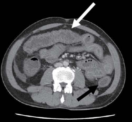
Figure 1 Abdominal CT scan showed diffuse edematous thickening of whole small bowel (white arrow) and colon with infammation (black arrow).

Figure 2 1×0.5 cm sized dark brown-colored eschar is shown in the right axillary area.
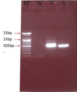
Figure 3 Gel electrophoresis of amplified DNAs by nested polymerase chain reaction of white cell buffy coat, which purified whole blood taken on day 4 showed band of 483 bp. M, 100bp DNA ladder; N, negative control; P, positive control; S, sample(pt)
Reference
-
1. Ogawa M, Hagiwara T, Kishimoto T, Shiga S, Yoshida Y, Furuya Y, Kaiho I, Ito T, Nemoto H, Yamamoto N, Masukawa K. Scrub typhus in Japan: epidemiology and clinical features of cases reported in 1998. Am J Trop Med Hyg. 2002. 67:162–165.
Article2. Kim DM, Kim SW, Choi SH, Yun NR. Clinical and laboratory findings associated with severe scrub typhus. BMC Infect Dis. 2010. 10:108.
Article3. Kim SJ, Chung IK, Chung IS, Song DH, Park SH, Kim HS, Lee MH. The clinical significance of upper gastrointestinal endoscopy in gastrointestinal vasculitis related to scrub typhus. Endoscopy. 2000. 32:950–955.
Article4. Hwang JH, Lee CS. Risk Factors Leading to Fatal Outcome in Scrub Typhus. JK Sci. 2010. 12:67–69.5. Lee YM, Kim DM, Lee SH, Jang MS, Neupane GP. Phylogenetic analysis of the 56 kDa protein genes of Orientia tsutsugamushi in southwest area of Korea. Am J Trop Med Hyg. 2011. 84:250–254.
Article6. Kim DM. Clinical features and diagnosis of scrub typhus. Infect Chemother. 2009. 41:315–322.
Article7. Lee N, Ip M, Wong B, Lui G, Tsang OT, Lai JY, Choi KW, Lam R, Ng TK, Ho J, Chan YY, Cockram CS, Lai ST. Risk factors associated with life-threatening rickettsial infections. Am J Trop Med Hyg. 2008. 78:973–978.
Article8. Balthazar EJ. CT of the gastrointestinal tract: principles and interpretation. AJR Am J Roentgenol. 1991. 156:23–32.
Article9. Macari M, Balthazar EJ. CT of bowel wall thickening: significance and pitfalls of interpretation. AJR Am J Roentgenol. 2001. 176:1105–1116.10. Nakayama K, Kurokawa K, Fukuhara M, Urakami H, Yamamoto S, Yamazaki K, Ogura Y, Ooka T, Hayashi T. Genome comparison and phylogenetic analysis of Orientia tsutsugamushi strains. DNA Res. 2010. 17:281–291.
Article11. Kim ES, Kim MK, Lee HM, Kil SH, Chung MH, Lee JS, Kang JS. Doxycycline resistance in Orientia tsutsugamushi isolated from Korean patients. Infect Chemother. 2008. 40:259–265.
Article12. Joo K, Kim MK, Kil SH, Chung MH, Kim JM, Kang JS. Cholestatic hepatitis caused by Tongyeong strain of Orientia tsutsugamushi. Infect Chemother. 2009. 41:99–104.
Article13. Park SW, Lee CK, Kwak YG, Moon C, Kim BN, Kim ES, Kang JM, Lee CS. Antigenic drift of Orientia tsutsugamushi in South Korea as identified by the sequence analysis of a 56-kDa protein-encoding gene. Am J Trop Med Hyg. 2010. 83:930–935.
Article14. Nakayama K, Yamashita A, Kurokawa K, Morimoto T, Ogawa M, Fukuhara M, Urakami H, Ohnishi M, Uchiyama I, Ogura Y, Ooka T, Oshima K, Tamura A, Hattori M, Hayashi T. The Whole-genome sequencing of the obligate intracellular bacterium Orientia tsutsugamushi revealed massive gene amplification during reductive genome evolution. DNA Res. 2008. 15:185–199.
Article15. Kelly DJ, Fuerst PA, Ching WM, Richards AL. Scrub typhus: the geographic distribution of phenotypic and genotypic variants of Orientia tsutsugamushi. Clin Infect Dis. 2009. 48:Suppl 3. S203–S230.
- Full Text Links
-
- Actions
-
Cited
- CITED
-
- Close
- Share
-
- Similar articles
-
- Severe Scrub Typhus Complicated by Myofascitis Due to the Taguchi Strain of Orientia tsutsugamushi
- Pulmonary Artery Thrombosis Associated with Scrub Typhus
- Cellular and Systemic Interactions of Orientia tsutsugamushi with Mammalian Host
- A Review of Electrocardiography Changes and Clinical Manifestations in Scrub Typhus in a Single Center
- Leukocytoclastic Vasculitis Associated with Orientia tsutsugamushi Infection: A Report of Two Cases
