Cellular and Systemic Interactions of Orientia tsutsugamushi with Mammalian Host
- Affiliations
-
- 1Department of Microbiology and Immunology, Seoul National University College of Medicine, Seoul, Korea. chonh@snu.ac.kr
- KMID: 2168662
- DOI: http://doi.org/10.4167/jbv.2012.42.4.276
Abstract
- Scrub typhus is an acute febrile illness caused by Orientia tsutsugamushi infection and one of main causes of febrile illness in the Asia-Pacific region. It has been estimated that one billion people are at risk and one million new cases arise each year in the endemic region. Despite of aggressive attempts to develop a prophylactic vaccine against scrub typhus during last several decades, all approaches have failed to generate long lasting immunity. In addition, little is known about the immunological pathogenesis of scrub typhus. In this review, we summarized recent findings of cellular and systemic interaction of O. tsutsugamushi with mammalian host, especially focusing on the molecular basis of intracellular invasion and immunological changes observed during the bacterial infection.
Figure
-
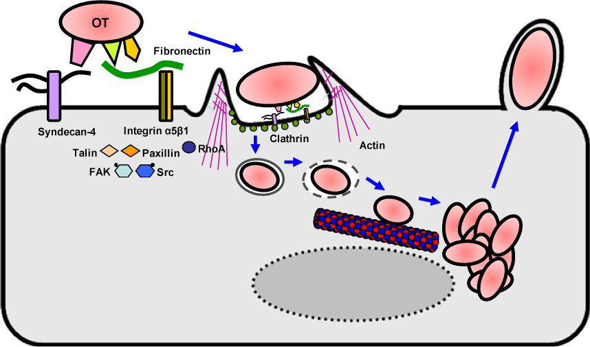
Figure 1 Hypothetical model for the intracellular invasion of O. tsutsugamushi. O. tsutsugamushi (OT) surface molecules (e.g., TSA56 and ScaC) mediate bacterial adhesion to host cells by binding to host-specific receptors, such as Syndecan-4 and fibronectin. The interaction between O. tsutsugamushi and fibronectin may mediate the engagement of integrins, which subsequently may activate downstream signaling molecules, such as FAK, Src kinase, and RhoA GTPase. Signaling adapters, such as talin and paxillin, are recruited to the site of infection. These signaling events consequently induce the internalization of O. tsutsugamushi into non-phagocytic host cells, which involves the clathrin-mediated endocytic pathway. The bacteria subsequently travel through the early and late endosomes and escape from the endosomal compartment within 2 h after infection. Cytosolic O. tsutsugamushi might move to microtubule organizing center via trafficking thorough microtubules and replicates in the perinuclear region (24).
Reference
-
1. Seong SY, Choi MS, Kim IS. Orientia tsutsugamushi infection: overview and immune responses. Microbes Infect. 2001. 3:11–21.2. Kelly DJ, Fuerst PA, Ching WM, Richards AL. Scrub typhus: the geographic distribution of phenotypic and genotypic variants of Orientia tsutsugamushi. Clin Infect Dis. 2009. 48:S203–S230.3. Watt G, Parola P. Scrub typhus and tropical rickettsioses. Curr Opin Infect Dis. 2003. 16:429–436.
Article4. Kweon SS, Choi JS, Lim HS, Kim JR, Kim KY, Ryu SY, et al. Rapid increase of scrub typhus, South Korea, 2001-2006. Emerg Infect Dis. 2009. 15:1127–1129.
Article5. Singh SI, Devi KP, Tilotama R, Ningombam S, Gopalkrishna Y, Singh TB, et al. An outbreak of scrub typhus in Bishnupur district of Manipur, India, 2007. Trop Doct. 2010. 40:169–170.
Article6. Zhang L, Jin Z, Xia S, Zhang J, Li M, Fu X, et al. Follow-up analysis on the epidemic strains of Orientia tsutsugamushi in the first outbreak of scrub typhus in Henan Province, China. Southeast Asian J Trop Med Public Health. 2007. 38:482–486.7. Zhang S, Song H, Liu Y, Li Q, Wang Y, Wu J, et al. Scrub typhus in previously unrecognized areas of endemicity in China. J Clin Microbiol. 2010. 48:1241–1244.
Article8. Izzard L, Fuller A, Blacksell SD, Paris DH, Richards AL, Aukkanit N, et al. Isolation of a novel Orientia species (O. chuto sp. nov.) from a patient infected in Dubai. J Clin Microbiol. 2010. 48:4404–4409.
Article9. Hendershot EF, Sexton DJ. Scrub typhus and rickettsial diseases in international travelers: a review. Curr Infect Dis Rep. 2009. 11:66–72.
Article10. Koh GC, Maude RJ, Paris DH, Newton PN, Blacksell SD. Diagnosis of scrub typhus. Am J Trop Med Hyg. 2010. 82:368–370.
Article11. Kelly DJ, Richards AL, Temenak J, Strickman D, Dasch GA. The past and present threat of rickettsial diseases to military medicine and international public health. Clin Infect Dis. 2002. 34:S145–S169.
Article12. Chattopadhyay S, Richards AL. Scrub typhus vaccines: past history and recent developments. Hum Vaccin. 2007. 3:73–80.
Article13. Kim DM, Won KJ, Park CY, Yu KD, Kim HS, Yang TY, et al. Distribution of eschars on the body of scrub typhus patients: a prospective study. Am J Trop Med Hyg. 2007. 76:806–809.
Article14. Jeong YJ, Kim S, Wook YD, Lee JW, Kim KI, Lee SH. Scrub typhus: clinical, pathologic, and imaging findings. Radiographics. 2007. 27:161–172.
Article15. Moron CG, Popov VL, Feng HM, Wear D, Walker DH. Identification of the target cells of Orientia tsutsugamushi in human cases of scrub typhus. Mod Pathol. 2001. 14:752–759.
Article16. Paris DH, Phetsouvanh R, Tanganuchitcharnchai A, Jones M, Jenjaroen K, Vongsouvath M, et al. Orientia tsutsugamushi in human scrub typhus eschars shows tropism for dendritic cells and monocytes rather than endothelium. PLoS Negl Trop Dis. 2012. 6:e1466.17. Cho NH, Seong SY, Huh MS, Han TH, Koh YS, Choi MS, et al. Expression of chemokine genes in murine macrophages infected with Orientia tsutsugamushi. Infect Immun. 2000. 68:594–602.
Article18. Rikihisa Y, Ito S. Localization of electron-dense tracers during entry of Rickettsia tsutsugamushi into polymorphonuclear leukocytes. Infect Immun. 1980. 30:231–243.
Article19. Cho NH, Seong SY, Choi MS, Kim IS. Expression of chemokine genes in human dermal microvascular endothelial cell lines infected with Orientia tsutsugamushi. Infect Immun. 2001. 69:1265–1272.
Article20. Cho BA, Cho NH, Seong SY, Choi MS, Kim IS. Intracellular invasion by Orientia tsutsugamushi is mediated by integrin signaling and actin cytoskeleton rearrangements. Infect Immun. 2010. 78:1915–1923.
Article21. Urakami H, Tsuruhara T, Tamura A. Penetration of Rickettsia tsutsugamushi into cultured mouse fibroblasts (L cells): an electron microscopic observation. Microbiol Immunol. 1983. 27:251–263.
Article22. Chu H, Lee JH, Han SH, Kim SY, Cho NH, Kim IS, et al. Exploitation of the endocytic pathway by Orientia tsutsugamushi in nonprofessional phagocytes. Infect Immun. 2006. 74:4246–4253.
Article23. Kim SW, Ihn KS, Han SH, Seong SY, Kim IS, Choi MS. Microtubule- and dynein-mediated movement of Orientia tsutsugamushi to the microtubule organizing center. Infect Immun. 2001. 69:494–500.
Article24. Ge Y, Rikihisa Y. Subversion of host cell signaling by Orientia tsutsugamushi. Microbes Infect. 2011. 13:638–648.25. Ihn KS, Han SH, Kim HR, Huh MS, Seong SY, Kang JS, et al. Cellular invasion of Orientia tsutsugamushi requires initial interaction with cell surface heparan sulfate. Microb Pathog. 2000. 28:227–233.
Article26. Kim HR, Choi MS, Kim IS. Role of Syndecan-4 in the cellular invasion of Orientia tsutsugamushi. Microb Pathog. 2004. 36:219–225.27. Lee JH, Cho NH, Kim SY, Bang SY, Chu H, Choi MS, et al. Fibronectin facilitates the invasion of Orientia tsutsugamushi into host cells through interaction with a 56-kDa type-specific antigen. J Infect Dis. 2008. 198:250–257.
Article28. Ha NY, Cho NH, Kim YS, Choi MS, Kim IS. An autotransporter protein from Orientia tsutsugamushi mediates adherence to nonphagocytic host cells. Infect Immun. 2011. 79:1718–1727.
Article29. Walsh DS, Myint KS, Kantipong P, Jongsakul K, Watt G. Orientia tsutsugamushi in peripheral white blood cells of patients with acute scrub typhus. Am J Trop Med Hyg. 2001. 65:899–901.
Article30. Koo JE, Yun JH, Lee KH, Hyun JW, Kang HK, Jang WJ, et al. Activation of mitogen-activated protein kinases is involved in the induction of interferon beta gene in macrophages infected with Orientia tsutsugamushi. Microbiol Immunol. 2009. 53:123–129.
Article31. Yun JH, Koo JE, Koh YS. Mitogen-activated protein kinases are involved in tumor necrosis factor alpha production in macrophages infected with Orientia tsutsugamushi. Microbiol Immunol. 2009. 53:349–355.
Article32. Geng P, Jerrells TR. The role of tumor necrosis factor in host defense against scrub typhus rickettsiae. I. Inhibition of growth of Rickettsia tsutsugamushi, Karp strain, in cultured murine embryonic cells and macrophages by recombinant tumor necrosis factor-alpha. Microbiol Immunol. 1994. 38:703–711.
Article33. Koo JE, Hong HJ, Dearth A, Kobayashi KS, Koh YS. Intracellular Invasion of Orientia tsutsugamushi Activates Inflammasome in ASC-Dependent Manner. PLoS ONE. 2012. 7:e39042.34. Cho NH, Seong SY, Huh MS, Kim NH, Choi MS, Kim IS. Induction of the gene encoding macrophage chemoattractant protein 1 by Orientia tsutsugamushi in human endothelial cells involves activation of transcription factor activator protein 1. Infect Immun. 2002. 70:4841–4850.
Article35. Cho KA, Jun YH, Suh JW, Kang JS, Choi HJ, Woo SY. Orientia tsutsugamushi induced endothelial cell activation via the NOD1-IL-32 pathway. Microb Pathog. 2010. 49:95–104.
Article36. Cho NH, Kim HR, Lee JH, Kim SY, Kim J, Cha S, et al. The Orientia tsutsugamushi genome reveals massive proliferation of conjugative type IV secretion system and host-cell interaction genes. Proc Natl Acad Sci U S A. 2007. 104:7981–7986.
Article37. Min CK, Yang JS, Kim S, Choi MS, Kim IS, Cho NH. Genome-Based Construction of the Metabolic Pathways of Orientia tsutsugamushi and Comparative Analysis within the Rickettsiales Order. Comp Funct Genomics. 2008. 623145.38. Nacy CA, Osterman JV. Host defenses in experimental scrub typhus: role of normal and activated macrophages. Infect Immun. 1979. 26:744–750.
Article39. Jerrells TR, Osterman JV. Host defenses in experimental scrub typhus: delayed-type hypersensitivity responses of inbred mice. Infect Immun. 1982. 35:117–123.
Article40. Kodama K, Kawamura S, Yasukawa M, Kobayashi Y. Establishment and characterization of a T-cell line specific for Rickettsia tsutsugamushi. Infect Immun. 1987. 55:2490–2495.
Article41. de Fost M, Chierakul W, Pimda K, Dondorp AM, White NJ, Van der Poll T. Activation of cytotoxic lymphocytes in patients with scrub typhus. Am J Trop Med Hyg. 2005. 72:465–467.
Article42. Seong SY, Huh MS, Jang WJ, Park SG, Kim JG, Woo SG, et al. Induction of homologous immune response to Rickettsia tsutsugamushi Boryong with a partial 56-kilodalton recombinant antigen fused with the maltose-binding protein MBP-Bor56. Infect Immun. 1997. 65:1541–1545.
Article43. Saunders JP, Brown GW, Shirai A, Huxsoll DL. The longevity of antibody to Rickettsia tsutsugamushi in patients with confirmed scrub typhus. Trans R Soc Trop Med Hyg. 1980. 74:253–257.
Article44. Eisenberg GH Jr, Osterman JV. Experimental scrub typhus immunogens: gamma-irradiated and formalinized rickettsiae. Infect Immun. 1977. 15:124–131.
Article45. Iwasaki H, Mizoguchi J, Takada N, Tai K, Ikegaya S, Ueda T. Correlation between the concentrations of tumor necrosis factor-alpha and the severity of disease in patients infected with Orientia tsutsugamushi. Int J Infect Dis. 2010. 14:e328–e333.46. Kramme S, An le V, Khoa ND, Trin le V, Tannich E, Rybniker J, et al. Orientia tsutsugamushi bacteremia and cytokine levels in Vietnamese scrub typhus patients. J Clin Microbiol. 2009. 47:586–589.
Article47. Iwasaki H, Takada N, Nakamura T, Ueda T. Increased levels of macrophage colony-stimulating factor, gamma interferon, and tumor necrosis factor alpha in sera of patients with Orientia tsutsugamushi infection. J Clin Microbiol. 1997. 35:3320–3322.
Article48. Chung DR, Lee YS, Lee SS. Kinetics of inflammatory cytokines in patients with scrub typhus receiving doxycycline treatment. J Infect. 2008. 56:44–50.
Article49. Sonthayanon P, Chierakul W, Wuthiekanun V, Phimda K, Pukrittayakamee S, Day NP, et al. Association of high Orientia tsutsugamushi DNA loads with disease of greater severity in adults with scrub typhus. J Clin Microbiol. 2009. 47:430–434.
Article50. Bourgeois AL, Olson JG, Fang RC, Huang J, Wang CL, Chow L, et al. Humoral and cellular responses in scrub typhus patients reflecting primary infection and reinfection with Rickettsia tsutsugamushi. Am J Trop Med Hyg. 1982. 31:532–540.
Article51. Ikeda M, Takahashi H, Yoshida S. HLA-DR+CD3+ and CD8+ cells are increased but CD4+CD45RA+ cells are reduced in the peripheral blood in human scrub typhus. Clin Immunol Immunopathol. 1994. 72:402–404.52. Cho BA, Ko Y, Kim YS, Kim S, Choi MS, Kim IS, et al. Phenotypic Characterization of Peripheral T cells and Their Dynamics in Scrub Typhus Patients. PLoS Negl Trop Dis. 2012. 6:e1789.
Article53. Chattopadhyay S, Jiang J, Chan TC, Manetz TS, Chao CC, Ching WM, et al. Scrub typhus vaccine candidate Kp r56 induces humoral and cellular immune responses in cynomolgus monkeys. Infect Immun. 2005. 73:5039–5047.
Article54. Jerrells TR. Immunosuppression associated with the development of chronic infections with Rickettsia tsutsugamushi: adherent suppressor cell activity and macrophage activation. Infect Immun. 1985. 50:175–182.
Article
- Full Text Links
-
- Actions
-
Cited
- CITED
-
- Close
- Share
-
- Similar articles
-
- Infectivity of Orientia tsutsugamushi to Various Eukaryotic Cells and Their Cellular Invasion Mechanism
- The Attachment of Detergent-Extracted Outer Membrane Proteins of Orientia tsutsugamushi to the Host Cell Surface
- Leukocytoclastic Vasculitis Associated with Orientia tsutsugamushi Infection: A Report of Two Cases
- Stabilizing Microtubular Network Facilitates the Intracellular Growth of Orientia tsutsugamushi
- Cleavage of p65 Subunit of NF-kappaB by Orientia tsutsugamushi
