Prediction of age-related osteoporosis using fractal analysis on panoramic radiographs
- Affiliations
-
- 1Department of Oral and Maxillofacial Radiology, School of Dentistry, and Institute of Oral Bio Science, Chonbuk National University, Jeonju, Korea. kkj1512@jbnu.ac.kr
- KMID: 1806789
- DOI: http://doi.org/10.5624/isd.2012.42.4.231
Abstract
- PURPOSE
This study was performed to evaluate the trabecular pattern on panoramic radiographs to predict age-related osteoporosis in postmenopausal women.
MATERIALS AND METHODS
Thirty-one postmenopausal osteoporotic women and 25 postmenopausal healthy women between the ages of 50 and 88 were enrolled in this study. The bone mineral density (BMD) of the lumbar vertebrae and femur were calculated using dual-energy X-ray absorptiometry (DXA), and panoramic radiographs were obtained. Fractal dimension (FD) was measured using the box counting method from 560 regions of interest (51x51 pixels) in 6 sites on the panoramic radiographs. The relationships between age and BMD and between FD and BMD were assessed, and the intraobserver agreement was determined.
RESULTS
There was a significant difference in the FD values between the osteoporotic and normal groups (p<0.05). There was a significant difference in the FD values at three sites in the jaws (p<0.05). Age was significantly correlated with the BMD measurements, with an odds ratio of 1.25. However, the FD values were not significantly correlated with the BMD measurements, with an odds ratio of 0.000. The intraobserver agreement showed relatively higher correlation coefficients at the upper premolar, lower premolar, and lower anterior regions than the other sites.
CONCLUSION
Age was an important risk factor for predicting the presence of osteoporosis in postmenopausal women. The lower premolar region was the most appropriate site for evaluating the FD value on panoramic radiographs. However, further investigation might be needed to predict osteoporosis using an FD value on panoramic radiographs.
MeSH Terms
Figure
-
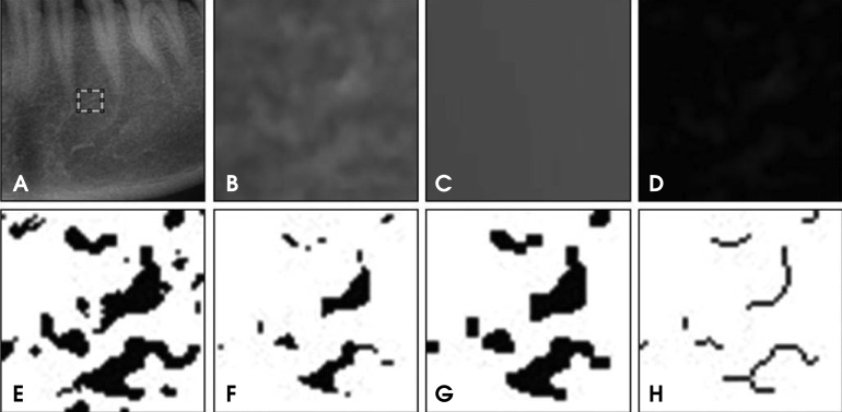
Fig. 1 Image processing procedures. A. The ROI (51×51 pixels) on a panoramic radiograph. B. The raw image before processing. C. A gaussian blurred image, D. A subtraction image. E. A binary image. F. An eroded image. G. A dilated image. H. A skeletonized image.
Cited by 5 articles
-
Method for Automated Selection of the Trabecular Area in Digital Periapical Radiographic Images Using Morphological Operations
Enny Itje Sela, Reza Pulungan, Rini Widyaningrum, Rurie Ratna Shantiningsih
Healthc Inform Res. 2019;25(3):193-200. doi: 10.4258/hir.2019.25.3.193.The three-dimensional microstructure of trabecular bone: Analysis of site-specific variation in the human jaw bone
Jo-Eun Kim, Jae-Myung Shin, Sung-Ook Oh, Won-Jin Yi, Min-Suk Heo, Sam-Sun Lee, Soon-Chul Choi, Kyung-Hoe Huh
Imaging Sci Dent. 2013;43(4):227-233. doi: 10.5624/isd.2013.43.4.227.IDIOS: An innovative index for evaluating dental imaging-based osteoporosis screening indices
Imad Barngkgei, Esam Halboub, Abeer Abdulkareem Almashraqi, Razan Khattab, Iyad Al Haffar
Imaging Sci Dent. 2016;46(3):185-202. doi: 10.5624/isd.2016.46.3.185.Comparison of cone-beam computed tomography and digital panoramic radiography for detecting peri-implant alveolar bone changes using trabecular micro-structure analysis
Guldane Magat, Elif Oncu, Sevgi Ozcan, Kaan Orhan
J Korean Assoc Oral Maxillofac Surg. 2022;48(1):41-49. doi: 10.5125/jkaoms.2022.48.1.41.Radiographic changes of mandibular cortical bone in bisphosphonate drug holiday
Dae-Hoon Lee, Ja-In Seo, Seung-Il Song, Jeong-Keun Lee
J Korean Assoc Oral Maxillofac Surg. 2022;48(4):219-224. doi: 10.5125/jkaoms.2022.48.4.219.
Reference
-
1. Verheij JG, Geraets WG, van der Stelt PF, Horner K, Lindh C, Nicopoulou-Karayianni K, et al. Prediction of osteoporosis with dental radiographs and age. Dentomaxillofac Radiol. 2009; 38:431–437. PMID: 19767512.
Article2. Geraets WG, van der Stelt PF. Fractal properties of bone. Dentomaxillofac Radiol. 2000; 29:144–153. PMID: 10849540.
Article3. Yaşar F, Akgünlü F. The differences in panoramic mandibular indices and fractal dimension between patients with and without spinal osteoporosis. Dentomaxillofac Radiol. 2006; 35:1–9. PMID: 16421256.
Article4. Horner K, Karayianni K, Mitsea A, Berkas L, Mastoris M, Jacobs R, et al. The mandibular cortex on radiographs as a tool for osteoporosis risk assessment: the OSTEODENT Project. J Clin Densitom. 2007; 10:138–146. PMID: 17449308.
Article5. Karayianni K, Horner K, Mitsea A, Berkas L, Mastoris M, Jacobs R, et al. Accuracy in osteoporosis diagnosis of a combination of mandibular cortical width measurement on dental panoramic radiographs and a clinical risk index (OSIRIS): the OSTEODENT project. Bone. 2007; 40:223–229. PMID: 16979965.
Article6. Horner K, Devlin H, Alsop CW, Hodgkinson IM, Adams JE. Mandibular bone mineral density as a predictor of skeletal osteoporosis. Br J Radiol. 1996; 69:1019–1025. PMID: 8958019.
Article7. Taguchi A, Suei Y, Sanada M, Ohtsuka M, Nakamoto T, Sumida H, et al. Validation of dental panoramic radiography measures for identifying postmenopausal women with spinal osteoporosis. AJR Am J Roentgenol. 2004; 183:1755–1760. PMID: 15547223.
Article8. Nackaerts O, Jacobs R, Devlin H, Pavitt S, Bleyen E, Yan B, et al. Osteoporosis detection using intraoral densitometry. Dentomaxillofac Radiol. 2008; 37:282–287. PMID: 18606750.
Article9. Kim JY, Jung YH, Nah KS. Prediction of osteoporosis using fractal analysis on periapical and panoramic radiographs. Korean J Oral Maxillofac Radiol. 2008; 38:147–151.10. Ruttimann UE, Ship JA. The use of fractal geometry to quantitate bone structure from radiographs. J Dent Res. 1990; 69:287. (Abstr 1431).11. Jolley L, Majumdar S, Kapila S. Technical factors in fractal analysis of periapical radiographs. Dentomaxillofac Radiol. 2006; 35:393–397. PMID: 17082328.
Article12. White SC, Rudolph DJ. Alterations of the trabecular pattern of the jaws in patients with osteoporosis. Oral Surg Oral Med Oral Pathol Oral Radiol Endod. 1999; 88:628–635. PMID: 10556761.
Article13. Amer ME, Heo MS, Brooks SL, Benavides E. Anatomical variations of trabecular bone structure in intraoral radiographs using fractal and particles count analyses. Imaging Sci Dent. 2012; 42:5–12. PMID: 22474642.
Article14. WHO Scientific Group on Research on the Menopause in the 1990s. WHO technical report series 866: research on the menopause in the 1990s. 1996. Geneva: World Health Organization.15. Heo MS, Park KS, Lee SS, Choi SC, Koak JY, Heo SJ, et al. Fractal analysis of mandibular bony healing after orthognathic surgery. Oral Surg Oral Med Oral Pathol Oral Radiol Endod. 2002; 94:763–767. PMID: 12464904.
Article16. Otis LL, Hong JS, Tuncay OC. Bone structure effect on root resorption. Orthod Craniofac Res. 2004; 7:165–177. PMID: 15359503.
Article17. Prouteau S, Ducher G, Nanyan P, Lemineur G, Benhamou L, Courteix D. Fractal analysis of bone texture: a screening tool for stress fracture risk? Eur J Clin Invest. 2004; 34:137–142. PMID: 14764077.
Article18. Chen SK, Oviir T, Lin CH, Leu LJ, Cho BH, Hollender L. Digital imaging analysis with mathematical morphology and fractal dimension for evaluation of periapical lesions following endodontic treatment. Oral Surg Oral Med Oral Pathol Oral Radiol Endod. 2005; 100:467–472. PMID: 16182168.
Article19. Ergün S, Saraçoğlu A, Güneri P, Özpinar B. Application of fractal analysis in hyperthyroidism. Dentomaxillofac Radiol. 2009; 38:281–288. PMID: 19474255.20. Southard TE, Southard KA, Jakobsen JR, Hillis SL, Najim CA. Fractal dimension in radiographic analysis of alveolar process bone. Oral Surg Oral Med Oral Pathol Oral Radiol Endod. 1996; 82:569–576. PMID: 8936523.
Article21. Shrout MK, Potter BJ, Hildebolt CF. The effect of image variations on fractal dimension calculations. Oral Surg Oral Med Oral Pathol Oral Radiol Endod. 1997; 84:96–100. PMID: 9247959.
Article22. An BM, Heo MS, Lee SP, Lee SS, Choi SC, Park TW, et al. Effect of exposure time and image resolution on fractal dimension. Korean J Oral Maxillofac Radiol. 2002; 32:75–79.
- Full Text Links
-
- Actions
-
Cited
- CITED
-
- Close
- Share
-
- Similar articles
-
- Prediction of osteoporosis using fractal analysis on periapical and panoramic radiographs
- Prediction of osteoporosis using fractal analysis et cetera on panoramic radiographs
- Structural complexity of the craniofacial trabecular bone in multiple myeloma assessed by fractal analysis
- Prediction of osteoporosis using fractal analysis on periapical radiographs
- Evaluation of peri-implant bone using fractal analysis
