Clin Orthop Surg.
2014 Sep;6(3):329-335. 10.4055/cios.2014.6.3.329.
Mini-Open Suture Bridge Repair with Porcine Dermal Patch Augmentation for Massive Rotator Cuff Tear: Surgical Technique and Preliminary Results
- Affiliations
-
- 1Department of Orthopedic Surgery, Dongsan Medical Center, Keimyung University School of Medicine, Daegu, Korea. oscho5362@dsmc.or.kr
- 2Department of Radiology, Dongsan Medical Center, Keimyung University School of Medicine, Daegu, Korea.
- 3Department of Orthopedic Surgery, Hanmi Hospital, Daegu, Korea.
- KMID: 1794716
- DOI: http://doi.org/10.4055/cios.2014.6.3.329
Abstract
- BACKGROUND
The aim of this study was to describe the mini-open suture bridge technique with porcine dermal patch augmentation for massive rotator cuff tear and to assess preliminary clinical and radiological results.
METHODS
Five patients with massive rotator cuff tear for which it was not possible to restore the anatomical footprint underwent mini-open suture bridge repair using a porcine dermal patch. The patients' average age was 53.4 years (range, 45 to 57 years), and the average duration of follow-up was 20.6 months (range, 14 to 26 months). Patients were evaluated with preoperative and postoperative outcome measures, including a visual analog scale (VAS) for pain, the University of California Los Angeles (UCLA) score, and the American Shoulder and Elbow Surgeons (ASES) score. The structural integrity of repaired rotator cuffs was assessed by magnetic resonance imaging 6 months postoperatively.
RESULTS
The average VAS pain score, UCLA score, and ASES score improved from 6.8, 15.4, and 39.4 preoperatively to 0.8, 31.2, and 86.4 postoperatively (p = 0.041, 0.042, and 0.043, respectively). Magnetic resonance images obtained at an average of 8 months after surgery showed that four patients had intact repair integrity with graft incorporation. One patient had a re-tear with partial healing but still had a satisfactory clinical outcome. There was no intraoperative or postoperative complication in any patient.
CONCLUSIONS
Mini-open suture bridge repair with porcine dermal patch augmentation can be an option in young patients with high physical demands and massive rotator cuff tears for which it is not possible to restore the anatomical footprint.
MeSH Terms
Figure
-
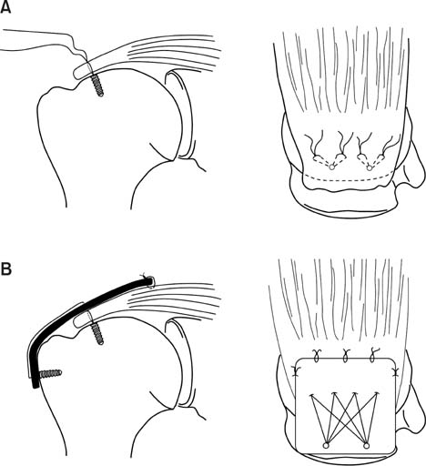
Fig. 1 Schematic illustrations of the mini-open suture bridge technique with porcine dermal patch augmentation. (A) Medialization of the footprint and medial row fixation using suture anchors. (B) Suture bridge fixation and marginal sutures with porcine dermal patch augmentation.
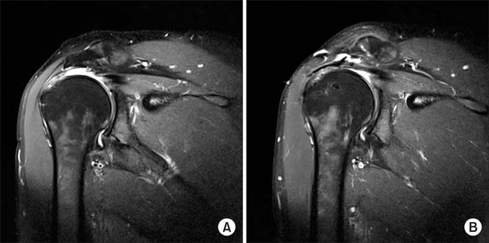
Fig. 2 Preoperative (A) and postoperative (B) magnetic resonance images. The image obtained 6 months after surgery shows an intact repair with graft incorporation.
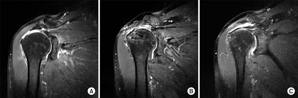
Fig. 3 Preoperative (A) and postoperative (B, C) magnetic resonance images. Images obtained 8 months after surgery show a full-thickness re-tear of the repaired supraspinatus (B) but with partial healing of the posterior edge of the rotator cuff and graft incorporation (C).
Reference
-
1. Bedi A, Dines J, Warren RF, Dines DM. Massive tears of the rotator cuff. J Bone Joint Surg Am. 2010; 92(9):1894–1908.2. Galatz LM, Ball CM, Teefey SA, Middleton WD, Yamaguchi K. The outcome and repair integrity of completely arthroscopically repaired large and massive rotator cuff tears. J Bone Joint Surg Am. 2004; 86(2):219–224.3. Kim JR, Cho YS, Ryu KJ, Kim JH. Clinical and radiographic outcomes after arthroscopic repair of massive rotator cuff tears using a suture bridge technique: assessment of repair integrity on magnetic resonance imaging. Am J Sports Med. 2012; 40(4):786–793.4. Mellado JM, Calmet J, Olona M, et al. Surgically repaired massive rotator cuff tears: MRI of tendon integrity, muscle fatty degeneration, and muscle atrophy correlated with intraoperative and clinical findings. AJR Am J Roentgenol. 2005; 184(5):1456–1463.5. Miller BS, Downie BK, Kohen RB, et al. When do rotator cuff repairs fail? Serial ultrasound examination after arthroscopic repair of large and massive rotator cuff tears. Am J Sports Med. 2011; 39(10):2064–2070.6. Oh JH, Kim SH, Shin SH, et al. Outcome of rotator cuff repair in large-to-massive tear with pseudoparalysis: a comparative study with propensity score matching. Am J Sports Med. 2011; 39(7):1413–1420.7. Papadopoulos P, Karataglis D, Boutsiadis A, Fotiadou A, Christoforidis J, Christodoulou A. Functional outcome and structural integrity following mini-open repair of large and massive rotator cuff tears: a 3-5 year follow-up study. J Shoulder Elbow Surg. 2011; 20(1):131–137.8. Ricchetti ET, Aurora A, Iannotti JP, Derwin KA. Scaffold devices for rotator cuff repair. J Shoulder Elbow Surg. 2012; 21(2):251–265.9. Rousseau T, Roussignol X, Bertiaux S, Duparc F, Dujardin F, Courage O. Arthroscopic repair of large and massive rotator cuff tears using the side-to-side suture technique: mid-term clinical and anatomic evaluation. Orthop Traumatol Surg Res. 2012; 98:4 Suppl. S1–S8.10. Domb BG, Glousman RE, Brooks A, Hansen M, Lee TQ, ElAttrache NS. High-tension double-row footprint repair compared with reduced-tension single-row repair for massive rotator cuff tears. J Bone Joint Surg Am. 2008; 90:Suppl 4. 35–39.11. Abrams JS, Song FS. Arthroscopic repair techniques for massive rotator cuff tears. Instr Course Lect. 2012; 61:121–130.12. Agrawal V. Healing rates for challenging rotator cuff tears utilizing an acellular human dermal reinforcement graft. Int J Shoulder Surg. 2012; 6(2):36–44.13. Bond JL, Dopirak RM, Higgins J, Burns J, Snyder SJ. Arthroscopic replacement of massive, irreparable rotator cuff tears using a GraftJacket allograft: technique and preliminary results. Arthroscopy. 2008; 24(4):403–409.e1.14. Coons DA, Alan Barber F. Tendon graft substitutes-rotator cuff patches. Sports Med Arthrosc. 2006; 14(3):185–190.15. Encalada-Diaz I, Cole BJ, Macgillivray JD, et al. Rotator cuff repair augmentation using a novel polycarbonate polyurethane patch: preliminary results at 12 months' follow-up. J Shoulder Elbow Surg. 2011; 20(5):788–794.16. Malcarney HL, Bonar F, Murrell GA. Early inflammatory reaction after rotator cuff repair with a porcine small intestine submucosal implant: a report of 4 cases. Am J Sports Med. 2005; 33(6):907–911.17. Xu H, Sandor M, Qi S, et al. Implantation of a porcine acellular dermal graft in a primate model of rotator cuff repair. J Shoulder Elbow Surg. 2012; 21(5):580–588.18. Baker AR, McCarron JA, Tan CD, Iannotti JP, Derwin KA. Does augmentation with a reinforced fascia patch improve rotator cuff repair outcomes? Clin Orthop Relat Res. 2012; 470(9):2513–2521.19. Ide J, Kikukawa K, Hirose J, Iyama K, Sakamoto H, Mizuta H. Reconstruction of large rotator-cuff tears with acellular dermal matrix grafts in rats. J Shoulder Elbow Surg. 2009; 18(2):288–295.20. Harper C. Permacol: clinical experience with a new biomaterial. Hosp Med. 2001; 62(2):90–95.21. Badhe SP, Lawrence TM, Smith FD, Lunn PG. An assessment of porcine dermal xenograft as an augmentation graft in the treatment of extensive rotator cuff tears. J Shoulder Elbow Surg. 2008; 17:1 Suppl. 35S–39S.22. Kim IB, Kim DJ. Arthroscopic xenograft repair of massive, irreparable rotator cuff tears. Clin Should Elbow. 2011; 14(2):214–221.23. Proper SI, Aladin A, Lam K, Lunn PG. Evaluation of a porcine dermal xenograft (PDX) in the treatment of chronic, massive rotator cuff defects. J Bone Joint Surg Br. 2003; 85:Suppl I. 69.24. Soler JA, Gidwani S, Curtis MJ. Early complications from the use of porcine dermal collagen implants (Permacol) as bridging constructs in the repair of massive rotator cuff tears: a report of 4 cases. Acta Orthop Belg. 2007; 73(4):432–436.25. Gerber C, Fuchs B, Hodler J. The results of repair of massive tears of the rotator cuff. J Bone Joint Surg Am. 2000; 82(4):505–515.26. Goutallier D, Postel JM, Bernageau J, Lavau L, Voisin MC. Fatty muscle degeneration in cuff ruptures: pre- and postoperative evaluation by CT scan. Clin Orthop Relat Res. 1994; (304):78–83.27. Cho CH, Song KS, Min BW, Jung GH, Lee YK, Sin HK. Anterolateral approach for mini-open rotator cuff repair. Int Orthop. 2012; 36(1):95–100.28. Rhee YG, Cho NS, Lim CT, Yi JW, Vishvanathan T. Bridging the gap in immobile massive rotator cuff tears: augmentation using the tenotomized biceps. Am J Sports Med. 2008; 36(8):1511–1518.29. Longo UG, Lamberti A, Rizzello G, Maffulli N, Denaro V. Synthetic augmentation in massive rotator cuff tears. Med Sport Sci. 2012; 57:168–177.30. Burkhart SS. Arthroscopic treatment of massive rotator cuff tears: clinical results and biomechanical rationale. Clin Orthop Relat Res. 1991; (267):45–56.
- Full Text Links
-
- Actions
-
Cited
- CITED
-
- Close
- Share
-
- Similar articles
-
- Anterolateral Mini-open Fixation with a Patch Augmentation for Latissimus Dorsi Tendon Transfer in Irreparable Rotator Cuff Tears: Technical Note
- Patch Augmentation for Massive Rotator Cuff Tears
- Reverse Total Shoulder Arthroplasty in the Massive Rotator Cuff Tear
- Arthroscopic Rotator Cuff Repair: Double Rows & Suture Bridge Technique
- Clinical Outcomes of Diverse Patch Grafts
