Incidental Findings on Knee Radiographs in Children and Adolescents
- Affiliations
-
- 1Department of Orthopedic Surgery, Seoul National University Bundang Hospital, Seongnam, Korea.
- 2Department of Orthopaedic Surgery, Myongji Hospital, Goyang, Korea. skh1219@naver.com
- 3Biomedical Research Institute, Seoul National University Bundang Hospital, Seongnam, Korea.
- 4Department of Orthopedic Surgery, Inha University Hospital, Incheon, Korea.
- 5Department of Orthopedic Surgery, Seoul National University Children's Hospital, Seoul, Korea.
- KMID: 1794712
- DOI: http://doi.org/10.4055/cios.2014.6.3.305
Abstract
- BACKGROUND
Despite the wide use of knee radiography in children and adolescent patients visiting the outpatient clinic, there has been no analysis about the prevalence and type of incidental findings yet. This study was performed to investigate the incidental findings on knee radiographs in children and adolescents according to age.
METHODS
A total of 1,562 consecutive patients younger than 18 years of age were included. They who visited Seoul National University Bundang Hospital's outpatient clinic with a chief complaint of knee pain or malalignment between 2010 and 2011. We reviewed the knee radiographs and analyzed the prevalence and type of incidental findings, such as metaphyseal lucent area, epiphyseal cortical irregularity, osteochondroma and Harris growth arrest line.
RESULTS
The mean age of the patients was 10.2 years (range, 1 month to 18 years). We identified 355 incidental findings in 335 patients (21.4%) and 98 abnormal findings (6.3%). The most common incidental finding was metaphyseal lucent area (131, 8.4%), followed by epiphyseal cortical irregularity (105, 6.7%), Harris growth arrest line (75, 4.8%), and osteochondroma (44, 2.8%). An epiphyseal cortical irregularity tended to have a higher prevalence at younger age (p < 0.001) and the prevalences of metaphyseal lucent area and Harris growth arrest line were also higher at a younger age (p = 0.001 and p < 0.001, respectively). However, the osteochondroma tended to have a higher prevalence at an older age (p = 0.004).
CONCLUSIONS
This study describes the incidental findings on knee radiographs in children and adolescents and provides effective information from a viewpoint of an orthopedic doctor. The authors recommend considering those incidental findings if unfamiliar findings appear on a knee radiograph in the pediatric outpatient clinic.
Keyword
MeSH Terms
Figure
-
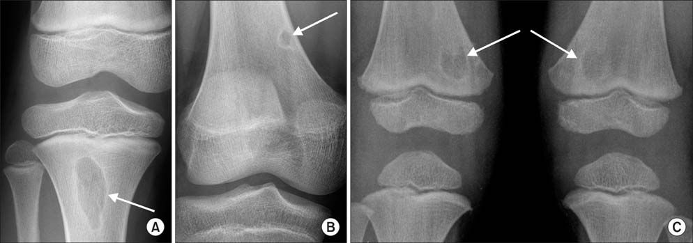
Fig. 1 Metaphyseal lucent area. (A) The non-ossifying fibroma is an enlarged benign cortical defect occurring in long bone of the lower extremity. (B) The cortical fibrous defect is a well-defined, intracortical, less than 2 cm round or oval radiolucency with sclerotic margins in the metaphyseal long bone. (C) Bilateral metaphyseal lucency refers to the presence of metaphyseal lucent areas in both lower extremities.
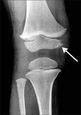
Fig. 2 Epiphyseal cortical irregularity showing a fragmented appearance on the medial side of the epiphysis in the distal femur.
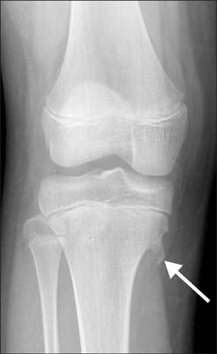
Fig. 3 Osteochondroma is a benign tumor characterized by an overgrowth of cartilage and bone near the end of the bone and near the growth plate.

Fig. 4 The Harris growth arrest line is an increased bone density representing the position of the growth plate.
Reference
-
1. Perquin CW, Hazebroek-Kampschreur AA, Hunfeld JA, et al. Pain in children and adolescents: a common experience. Pain. 2000; 87(1):51–58.2. Goodman JE, McGrath PJ. The epidemiology of pain in children and adolescents: a review. Pain. 1991; 46(3):247–264.3. Baena-Ocampo Ldel C, Ramirez-Perez E, Linares-Gonzalez LM, Delgado-Chavez R. Epidemiology of bone tumors in Mexico City: retrospective clinicopathologic study of 566 patients at a referral institution. Ann Diagn Pathol. 2009; 13(1):16–21.4. Solooki S, Vosoughi AR, Masoomi V. Epidemiology of musculoskeletal tumors in Shiraz, south of Iran. Indian J Med Paediatr Oncol. 2011; 32(4):187–191.5. Walden MJ, Murphey MD, Vidal JA. Incidental enchondromas of the knee. AJR Am J Roentgenol. 2008; 190(6):1611–1615.6. Simon H. Medial distal metaphyseal femoral irregularity in children. Radiology. 1968; 90(2):258–260.7. Vieira RL, Bencardino JT, Rosenberg ZS, Nomikos G. MRI features of cortical desmoid in acute knee trauma. AJR Am J Roentgenol. 2011; 196(2):424–428.8. Johnson LC, Genner BA III, Engh CA, Brown RH. Cortical desmoids. J Bone Joint Surg Am. 1968; 50(4):828–829.9. Bufkin WJ. The avulsive cortical irregularity. Am J Roentgenol Radium Ther Nucl Med. 1971; 112(3):487–492.10. Sklar DH, Phillips JJ, Lachman RS. Case report 683: distal metaphyseal femoral defect (cortical desmoid; distal femoral cortical irregularity). Skeletal Radiol. 1991; 20(5):394–396.11. Betsy M, Kupersmith LM, Springfield DS. Metaphyseal fibrous defects. J Am Acad Orthop Surg. 2004; 12(2):89–95.12. Swischuk LE. Imaging of the new bone, infant and young child. 5th ed. Philadelphia, PA: Lippincott Williams & Wikins;2003. p. 725–734.13. Verdonk PC, Verstraete K, Verdonk R. Distal femoral cortical irregularity in a 13-year old boy: a case report. Acta Orthop Belg. 2003; 69(4):377–381.14. Wolf SM, Paradise J, Caga-anan C. The law of incidental findings in human subjects research: establishing researchers' duties. J Law Med Ethics. 2008; 36(2):361–383.15. Kumar R, Madewell JE, Lindell MM, Swischuk LE. Fibrous lesions of bones. Radiographics. 1990; 10(2):237–256.16. Resnick D, Kransdorf MJ. Bone and joint imaging. 3rd ed. Philadelphia, PA: Elsevier Saunders;2005.17. Lange RH, Lange TA, Rao BK. Correlative radiographic, scintigraphic, and histological evaluation of exostoses. J Bone Joint Surg Am. 1984; 66(9):1454–1459.18. Ecklund K, Jaramillo D. Imaging of growth disturbance in children. Radiol Clin North Am. 2001; 39(4):823–841.19. Bonett DG. Sample size requirements for estimating intraclass correlations with desired precision. Stat Med. 2002; 21(9):1331–1335.20. Lee KM, Lee J, Chung CY, et al. Pitfalls and important issues in testing reliability using intraclass correlation coefficients in orthopaedic research. Clin Orthop Surg. 2012; 4(2):149–155.21. Choong PF, Pritchard DJ, Rock MG, Sim FH, McLeod RA, Unni KK. Low grade central osteogenic sarcoma: a long-term followup of 20 patients. Clin Orthop Relat Res. 1996; (322):198–206.22. Papathanassiou ZG, Alberghini M, Thiesse P, et al. Parosteal osteosarcoma mimicking osteochondroma: a radio-histologic approach on two cases. Clin Sarcoma Res. 2011; 1(1):2.23. Kristy LW, Mary IO. Benign cartilage tumors. In : Herbert SS, editor. Orthopaedic knowledge update: musculoskeletal tumors 2. Rosemont, IL: American Academy of Orthopaedic Surgeons;2007. p. 103–120.24. Subhas N, Bui KL, Sundaram M, Ilaslan H, Recht MP. Incidental tumor and tumor-like lesions around the knee. Semin Musculoskelet Radiol. 2009; 13(4):353–370.25. Kleinman PK, Belanger PL, Karellas A, Spevak MR. Normal metaphyseal radiologic variants not to be confused with findings of infant abuse. AJR Am J Roentgenol. 1991; 156(4):781–783.26. Tang CW, Kay RM, Skaggs DL. Growth arrest of the distal radius following a metaphyseal fracture: case report and review of the literature. J Pediatr Orthop B. 2002; 11(1):89–92.27. Carey J, Spence L, Blickman H, Eustace S. MRI of pediatric growth plate injury: correlation with plain film radiographs and clinical outcome. Skeletal Radiol. 1998; 27(5):250–255.28. Havranek P, Lizler J. Magnetic resonance imaging in the evaluation of partial growth arrest after physeal injuries in children. J Bone Joint Surg Am. 1991; 73(8):1234–1241.29. Sontag LW, Harris LM. Evidences of disturbed prenatal and neonatal growth in bones of infants aged one month. II. contributing factors. Am J Dis Child. 1938; 56(6):1248–1255.30. Gindhart PS. The frequency of appearance of transverse lines in the tibia in relation to childhood illnesses. Am J Phys Anthropol. 1969; 31(1):17–22.
