Differentiating Benign from Malignant Bone Tumors Using Fluid-Fluid Level Features on Magnetic Resonance Imaging
- Affiliations
-
- 1Department of Radiology, The Third Hospital of Hebei Medical University, Hebei Province Biomechanical Key Laborary of Orthopedics, Shijiazhuang, Hebei 050051, China. jianlingcui@sina.com
- KMID: 1794647
- DOI: http://doi.org/10.3348/kjr.2014.15.6.757
Abstract
OBJECTIVE
To analyze different fluid-fluid level features between benign and malignant bone tumors on magnetic resonance imaging (MRI).
MATERIALS AND METHODS
This study was approved by the hospital ethics committee. We retrospectively analyzed 47 patients diagnosed with benign (n = 29) or malignant (n = 18) bone tumors demonstrated by biopsy/surgical resection and who showed the intratumoral fluid-fluid level on pre-surgical MRI. The maximum length of the largest fluid-fluid level and the ratio of the maximum length of the largest fluid-fluid level to the maximum length of a bone tumor in the sagittal plane were investigated for use in distinguishing benign from malignant tumors using the Mann-Whitney U-test and a receiver operating characteristic (ROC) analysis. Fluid-fluid level was categorized by quantity (multiple vs. single fluid-fluid level) and by T1-weighted image signal pattern (high/low, low/high, and undifferentiated), and the findings were compared between the benign and malignant groups using the chi2 test.
RESULTS
The ratio of the maximum length of the largest fluid-fluid level to the maximum length of bone tumors in the sagittal plane that allowed statistically significant differentiation between benign and malignant bone tumors had an area under the ROC curve of 0.758 (95% confidence interval, 0.616-0.899). A cutoff value of 41.5% (higher value suggests a benign tumor) had sensitivity of 73% and specificity of 83%.
CONCLUSION
The ratio of the maximum length of the largest fluid-fluid level to the maximum length of a bone tumor in the sagittal plane may be useful to differentiate benign from malignant bone tumors.
MeSH Terms
-
Adolescent
Adult
Aged
Area Under Curve
Bone Neoplasms/diagnosis/*radiography/surgery
Child
Female
Humans
Image Processing, Computer-Assisted
*Magnetic Resonance Imaging
Male
Middle Aged
Precancerous Conditions/diagnosis/*radiography
ROC Curve
Retrospective Studies
Sensitivity and Specificity
Statistics, Nonparametric
Young Adult
Figure
-

Fig. 1 Receiver operating characteristic (ROC) curve when ratio of maximum length of largest fluid-fluid level to maximum length of bone tumor in sagittal plane is > 41.5%.
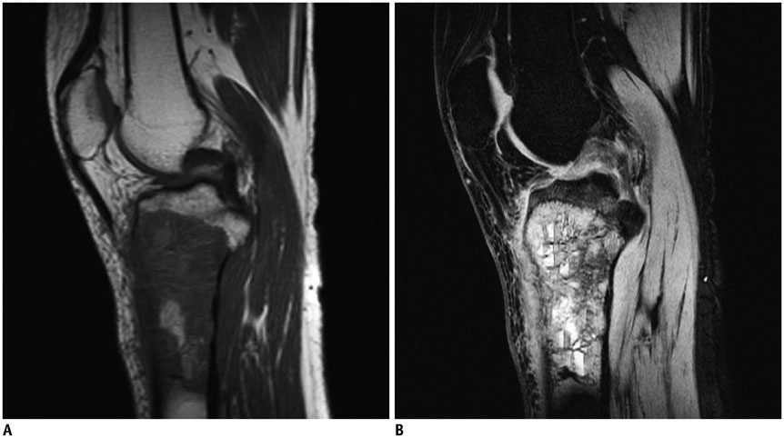
Fig. 2 Sagittal T1-weighted (A) and short tau inversion recovery (B) magnetic resonance images in 17-year-old male with osteosarcoma of proximal tibia. Fluid-fluid level cannot be found in A, but multiple fluid-fluid levels can be found in B, and ratio of maximum length of largest fluid-fluid level to maximum length of tumor was 13.8%. Fluid-fluid levels show high/low signal pattern in B.
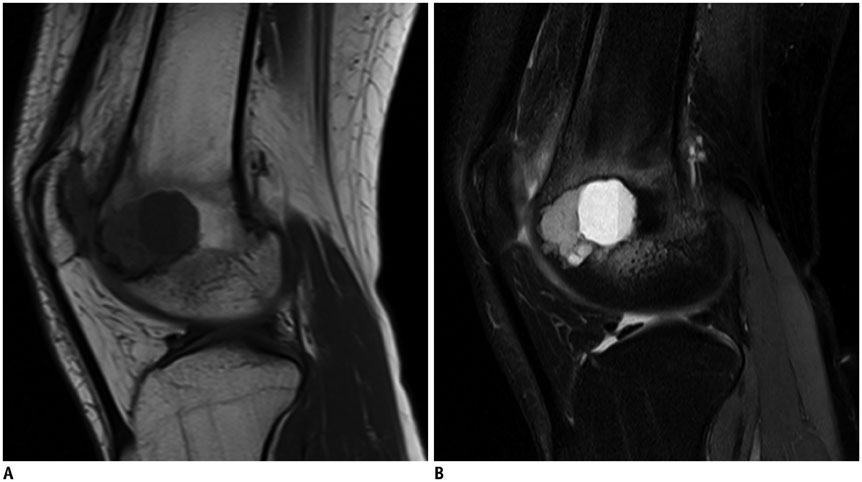
Fig. 3 Sagittal T1-weighted (A) and short tau inversion recovery (B) magnetic resonance images in 28-year-old male with chondroblastoma of distal femur. Tumor contained single fluid-fluid level, and ratio of maximum length of largest fluid-fluid level to maximum length of tumor was 81.8%. Fluid-fluid level shows low/high signal pattern in A and high/low in B.

Fig. 4 Sagittal T1-weighted (A) and short tau inversion recovery (B) magnetic resonance images in 43-year-old woman with aneurysmal bone cyst of proximal tibia. Lesion contains two fluid-fluid levels, and fluid-fluid levels show high/low signal pattern in A and B. Ratio of maximum length of largest fluid-fluid level to maximum length of lesion was about 61.2%.
Reference
-
1. Hudson TM. Fluid levels in aneurysmal bone cysts: a CT feature. AJR Am J Roentgenol. 1984; 142:1001–1004.2. Davies AM, Cassar-Pullicino VN, Grimer RJ. The incidence and significance of fluid-fluid levels on computed tomography of osseous lesions. Br J Radiol. 1992; 65:193–198.3. Tsai JC, Dalinka MK, Fallon MD, Zlatkin MB, Kressel HY. Fluid-fluid level: a nonspecific finding in tumors of bone and soft tissue. Radiology. 1990; 175:779–782.4. Burr BA, Resnick D, Syklawer R, Haghighi P. Fluid-fluid levels in a unicameral bone cyst: CT and MR findings. J Comput Assist Tomogr. 1993; 17:134–136.5. Buetow PC, Newman S, Kransdorf MJ. Giant-cell tumor of the tibia in a child presenting as an expansile metaphyseal lesion with fluid-fluid levels on MR. Magn Reson Imaging. 1990; 8:341–344.6. Fisher AJ, Totty WG, Kyriakos M. MR appearance of cystic fibrous dysplasia. J Comput Assist Tomogr. 1994; 18:315–318.7. Van Dyck P, Vanhoenacker FM, Vogel J, Venstermans C, Kroon HM, Gielen J, et al. Prevalence, extension and characteristics of fluid-fluid levels in bone and soft tissue tumors. Eur Radiol. 2006; 16:2644–2651.8. O'Donnell P, Saifuddin A. The prevalence and diagnostic significance of fluid-fluid levels in focal lesions of bone. Skeletal Radiol. 2004; 33:330–336.9. Alyas F, Saifuddin A. Fluid-fluid levels in bone neoplasms: variation of T1-weighted signal intensity of the superior to inferior layers--diagnostic significance on magnetic resonance imaging. Eur Radiol. 2008; 18:2642–2651.10. Lodwick GS, Wilson AJ, Farrell C, Virtama P, Dittrich F. Determining growth rates of focal lesions of bone from radiographs. Radiology. 1980; 134:577–583.11. Asaumi J, Konouchi H, Hisatomi M, Matsuzaki H, Shigehara H, Honda Y, et al. MR features of aneurysmal bone cyst of the mandible and characteristics distinguishing it from other lesions. Eur J Radiol. 2003; 45:108–112.12. Chung CB, Murphey M, Cho G, Schweitzer M, Hodler J, Haghihi P, et al. Osseous lesions of the pelvis and long tubular bones containing both fat and fluid-like signal intensity: an analysis of 28 patients. Eur J Radiol. 2005; 53:103–109.13. Hudson TM, Hamlin DJ, Fitzsimmons JR. Magnetic resonance imaging of fluid levels in an aneurysmal bone cyst and in anticoagulated human blood. Skeletal Radiol. 1985; 13:267–270.14. Nguyen BD, Westra WH, Kuhlman JE. Bone metastasis from breast carcinoma with fluid-fluid level. Skeletal Radiol. 1996; 25:189–192.
- Full Text Links
-
- Actions
-
Cited
- CITED
-
- Close
- Share
-
- Similar articles
-
- Evaluation of malignant intraductal papillary mucinous neoplasms of the pancreas on computed tomography and magnetic resonance imaging
- A Case Report of Multiple Cavernous Hemangiomas with Fluid-Fluid Levels: A Focus on the Radiologic Features of Dynamic MRI with Subtraction
- Imaging Findings of Sacral Tumors
- Cerebrospinal fluid flow in normal beagle dogs analyzed using magnetic resonance imaging
- MRI Findings and Differential Diagnosis of Benign and Malignant Tumors of the Uterine Corpus
