Korean J Urol.
2014 Aug;55(8):542-550. 10.4111/kju.2014.55.8.542.
Altered Gene Expression Profile After Exposure to Transforming Growth Factor beta1 in the 253J Human Bladder Cancer Cell Line
- Affiliations
-
- 1Department of Urology, Soonchunhyang University Cheonan Hospital, Cheonan, Korea.
- 2Department of Urology, Seoul National University College of Medicine, Seoul, Korea.
- 3Department of Biochemistry, Soonchunhyang University College of Medicine, Cheonan, Korea.
- 4Department of Urology, Seoul National University Bundang Hospital, Seongnam, Korea. selee@snubh.org
- KMID: 1794590
- DOI: http://doi.org/10.4111/kju.2014.55.8.542
Abstract
- PURPOSE
Transforming growth factor beta1 (TGF-beta1) inhibits the growth of bladder cancer cells and this effect is prominent and constant in 253J bladder cancer cells. We performed a microarray analysis to search for genes that were altered after TGF-beta1 treatment to understand the growth inhibitory action of TGF-beta1.
MATERIALS AND METHODS
253J bladder cancer cells were exposed to TGF-beta1 and total RNA was extracted at 6, 24, and 48 hours after exposure. The RNA was hybridized onto a human 22K oligonucleotide microarray and the data were analyzed by using GeneSpring 7.1.
RESULTS
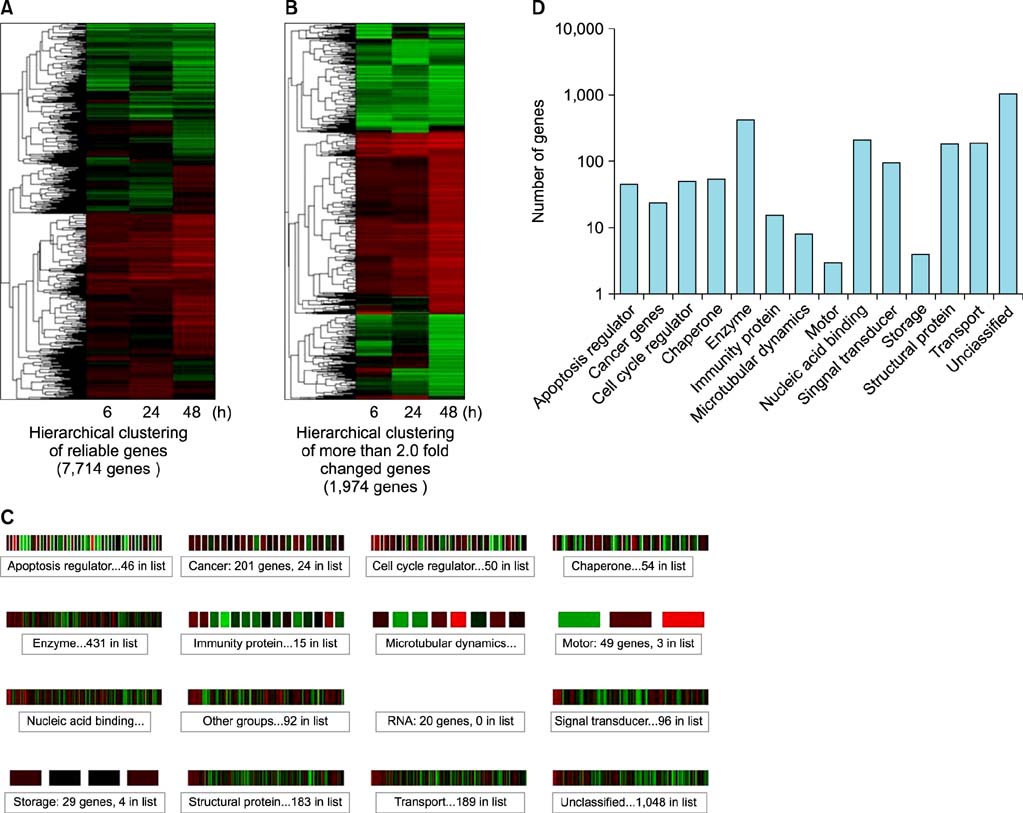
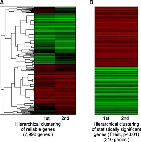
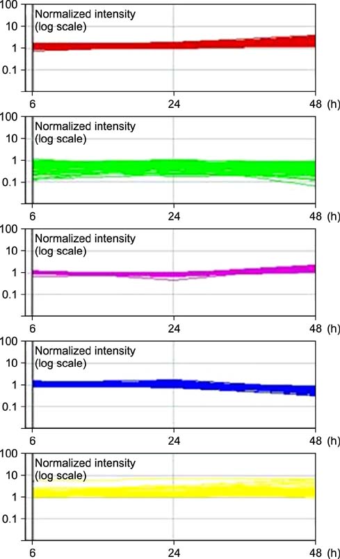
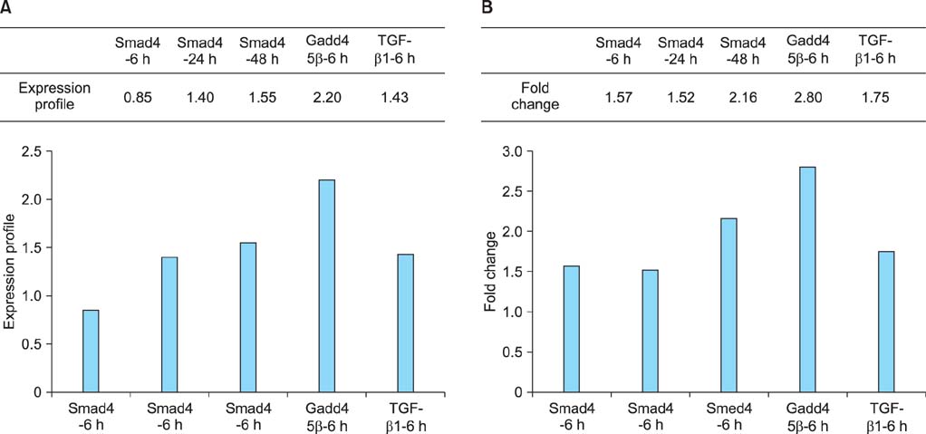
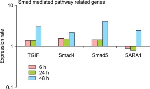
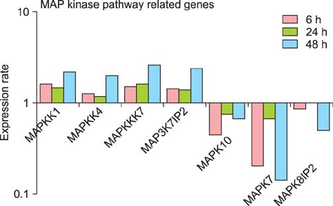
In the microarray analysis, a total of 1,974 genes showing changes of more than 2.0 fold were selected. The selected genes were further subdivided into five highly cohesive clusters with high probability according to the time-dependent expression pattern. A total of 310 genes showing changes of more than 2.0 fold in repeated arrays were identified by use of simple t-tests. Of these genes, those having a known function were listed according to clusters. Microarray analysis showed increased expression of molecules known to be related to Smad-dependent signal transduction, such as SARA and Smad4, and also those known to be related to the mitogen-activated protein kinase (MAPK) pathway, such as MAPKK1 and MAPKK4.
CONCLUSIONS
A list of genes showing significantly altered expression profiles after TGF-beta1 treatment was made according to five highly cohesive clusters. The data suggest that the growth inhibitory effect of TGF-beta1 in bladder cancer may occur through the Smad-dependent pathway, possibly via activation of the extracellular signal-related kinase 1 and Jun amino-terminal kinases Mitogen-activated protein kinase pathway.
Keyword
MeSH Terms
-
Antineoplastic Agents/*pharmacology
Cluster Analysis
Gene Expression Profiling/methods
Gene Expression Regulation, Neoplastic/*drug effects
Genes, Neoplasm
Humans
MAP Kinase Signaling System/drug effects/genetics
Neoplasm Proteins/genetics/metabolism
Oligonucleotide Array Sequence Analysis/methods
Reverse Transcriptase Polymerase Chain Reaction/methods
Signal Transduction/drug effects/genetics
Smad Proteins/genetics/metabolism
Transforming Growth Factor beta1/*pharmacology
Tumor Cells, Cultured/drug effects
Urinary Bladder Neoplasms/*genetics/metabolism/pathology
Antineoplastic Agents
Neoplasm Proteins
Smad Proteins
Transforming Growth Factor beta1
Figure
Reference
-
1. Massague J. TGF-beta signal transduction. Annu Rev Biochem. 1998; 67:753–791.2. Blobe GC, Schiemann WP, Lodish HF. Role of transforming growth factor beta in human disease. N Engl J Med. 2000; 342:1350–1358.3. Massague J, Blain SW, Lo RS. TGFbeta signaling in growth control, cancer, and heritable disorders. Cell. 2000; 103:295–309.4. Lee C, Lee SH, Kim DS, Jeon YS, Lee NK, Lee SE. Growth inhibition after exposure to transforming growth factor-β1 in human bladder cancer cell lines. Korean J Urol. 2014; 55:487–492.5. Rocke DM, Durbin B. A model for measurement error for gene expression arrays. J Comput Biol. 2001; 8:557–569.6. Yang YH, Dudoit S, Luu P, Lin DM, Peng V, Ngai J, et al. Normalization for cDNA microarray data: a robust composite method addressing single and multiple slide systematic variation. Nucleic Acids Res. 2002; 30:e15.7. Kerr G, Ruskin HJ, Crane M, Doolan P. Techniques for clustering gene expression data. Comput Biol Med. 2008; 38:283–293.8. Budhraja V, Spitznagel E, Schaiff WT, Sadovsky Y. Incorporation of gene-specific variability improves expression analysis using high-density DNA microarrays. BMC Biol. 2003; 1:1.9. Derynck R, Zhang YE. Smad-dependent and Smad-independent pathways in TGF-beta family signalling. Nature. 2003; 425:577–584.10. Sanchez-Capelo A. Dual role for TGF-beta1 in apoptosis. Cytokine Growth Factor Rev. 2005; 16:15–34.11. Tsukazaki T, Chiang TA, Davison AF, Attisano L, Wrana JL. SARA, a FYVE domain protein that recruits Smad2 to the TGFbeta receptor. Cell. 1998; 95:779–791.12. Govinden R, Bhoola KD. Genealogy, expression, and cellular function of transforming growth factor-beta. Pharmacol Ther. 2003; 98:257–265.13. Chang L, Karin M. Mammalian MAP kinase signalling cascades. Nature. 2001; 410:37–40.14. Yue J, Mulder KM. Activation of the mitogen-activated protein kinase pathway by transforming growth factor-beta. Methods Mol Biol. 2000; 142:125–131.
- Full Text Links
- Actions
-
Cited
- CITED
-
- Close
- Share
- Similar articles
-
- Growth Inhibition After Exposure to Transforming Growth Factor-beta1 in Human Bladder Cancer Cell Lines
- Inhibition of Cell Growth and Suppression of c-myc Gene Expression by Transforming Growth Factor-beta1 in Cervical Carcinoma Cell Lines
- Altered Expression of Genes in a Human Urinary Bladder Transitional Cell Carcinoma Cell Line of Low-metastatic Potential and Its Subclone of High-metastatic Potential
- The Transforming Growth Factor-beta1 Expression in Normal Laryngeal Mucosa, Laryngeal Dysplasia and Laryngeal Carcinoma
- Effects of Transforming Growth Factor-beta1 and Its Receptor on the Development, Recurrence and Progression of Human Bladder Cancer

