Effects of Argon Laser Iridotomy on the Corneal Endothelium of Pigmented Rabbit Eyes
- Affiliations
-
- 1Department of Ophthalmology, Korea University College of Medicine, Seoul, Korea. crisim@korea.ac.kr
- KMID: 1792096
- DOI: http://doi.org/10.3341/kjo.2014.28.1.76
Abstract
- PURPOSE
In Asian countries, laser iridotomy for the treatment of angle-closure glaucoma is a common cause of bullous keratopathy, which may be associated with a shallow anterior chamber and dark iris pigmentation in Asians. Several cases of corneal decompensation after argon laser iridotomy have been reported. In the present study, we evaluated the harmful effects of argon laser iridotomy on the corneal endothelium.
METHODS
Argon laser iridotomy was performed on the right eyes of pigmented rabbits. Changes in corneal thickness and endothelial cell density after laser iridotomy were evaluated. Terminal deoxynucleotidyl transferase dUTP nick end labeling (TUNEL) was performed for assessment of corneal endothelial cell apoptosis. Combined staining with alizarin red and trypan blue, as well as a live/dead cell assay, were performed for evaluation of damage to the corneal endothelium induced by laser iridotomy.
RESULTS
Corneal thickness did not change immediately after laser iridotomy; however, a significant increase was observed 24 hours after iridotomy (p = 0.001). The endothelial cell density of laser-treated eyes four days after laser iridotomy was significantly decreased compared with control eyes (p < 0.001). TUNEL staining showed many TUNEL-positive cells in the corneal endothelium and corneal stroma. No endothelial trypan blue-stained cell nuclei were observed after laser iridotomy; however, several large endothelial cells with damaged membrane integrity were observed. The live/dead cell assay clearly showed a large number of dead cells stained red in several areas throughout the entire corneal button 24 hours after iridotomy.
CONCLUSIONS
Argon laser iridotomy induces corneal endothelial cell apoptosis in pigmented rabbit eyes, resulting in decreased endothelial cell density.
Keyword
MeSH Terms
Figure
-
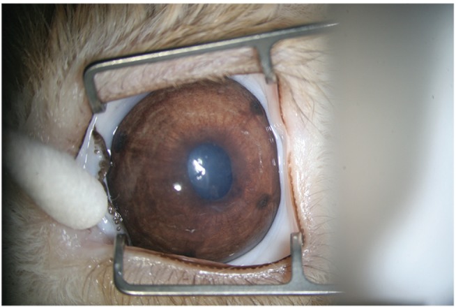
Fig. 1 A rabbit eye after argon laser peripheral iridotomy in each of four quadrants (superior temporal, superior nasal, inferior temporal, and inferior nasal) using an Abraham iridotomy contact lens.
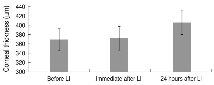
Fig. 2 Change in mean central corneal thickness after argon laser iridotomy (LI). Although corneal thickness did not differ significantly before and immediately after iridotomy (p = 0.276), it showed a significant increase 24 hours after iridotomy compared with before LI (p = 0.001, n = 10).
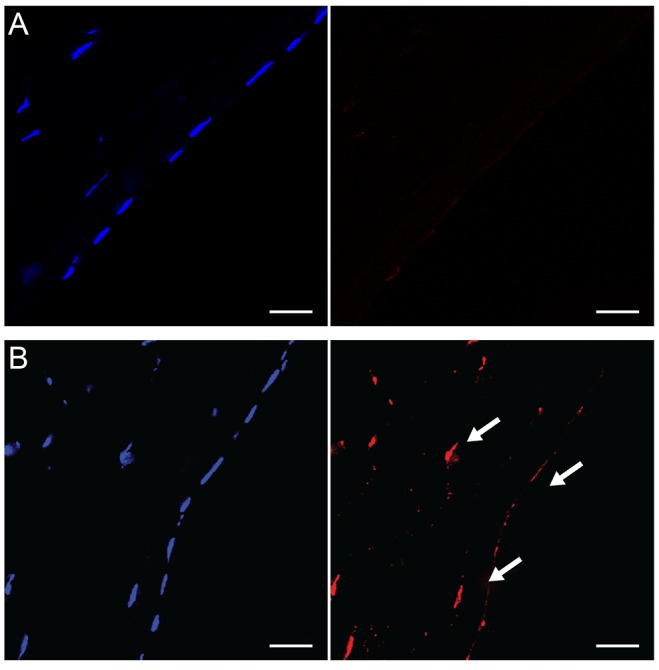
Fig. 3 (A) In normal rabbit corneas, terminal deoxynucleotidyl transferase dUTP nick end labeling (TUNEL)-positive cells were not detected in the corneal stroma or corneal endothelium. (B) Twenty-four hours after argon laser iridotomy, many TUNEL-positive cells (arrows) were observed in the corneal endothelium as well as in the corneal stroma. TUNEL staining, bar = 20 µm.
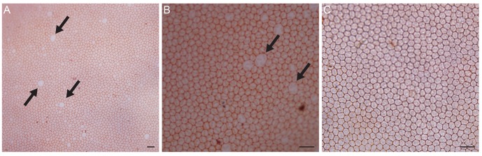
Fig. 4 (A) Twenty-four hours after iridotomy, intercellular borders of endothelial cells were clearly stained with alizarin red, and several large endothelial cells with damaged membrane integrity (arrows) were observed. (B) Endothelial cells with trypan blue-stained nuclei were not observed. (C) Four days after iridotomy, damaged endothelial cells were no longer found, and a normal mosaic pattern was observed. Dual staining of trypan blue and alizarin red, bar = 40 µm.
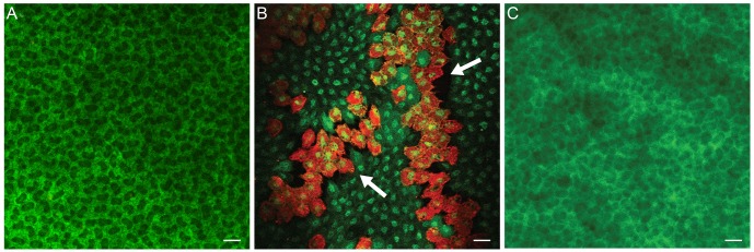
Fig. 5 (A) Almost all endothelial cells of normal cornea were viable and stained green. (B) One day after iridotomy, many dead cells stained red were observed in several areas throughout the corneal button. (C) Four days after iridotomy, dead cells were no longer observed. Live/dead cell assay, bar = 20 µm.
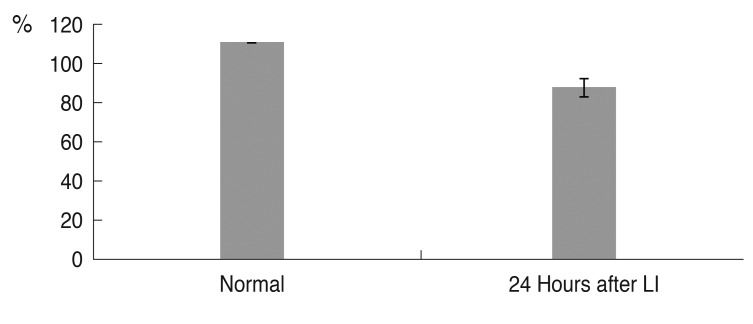
Fig. 6 The mean percentage of live cells in the corneal endothelium. Although 100% of corneal endothelial cells in the normal cornea were alive, 24 hours after laser iridotomy (LI), the percentage of live cells was 78.9% (n = 4, p < 0.001).
Reference
-
1. Murphy C, Alvarado J, Juster R, Maglio M. Prenatal and postnatal cellularity of the human corneal endothelium. A quantitative histologic study. Invest Ophthalmol Vis Sci. 1984; 25:312–322. PMID: 6698749.2. Joyce NC. Proliferative capacity of the corneal endothelium. Prog Retin Eye Res. 2003; 22:359–389. PMID: 12852491.
Article3. Klyce SD. Corneal physiology. In : Foster CS, Azar DT, Dohlman CH, editors. Smolin and Thoft's the cornea. Philadelphia: Lippincott Williams & Wilkins;2004. p. 37–58.4. Cho KS, Lee EH, Choi JS, Joo CK. Reactive oxygen species-induced apoptosis and necrosis in bovine corneal endothelial cells. Invest Ophthalmol Vis Sci. 1999; 40:911–919. PMID: 10102288.5. Koh SW, Waschek JA. Corneal endothelial cell survival in organ cultures under acute oxidative stress: effect of VIP. Invest Ophthalmol Vis Sci. 2000; 41:4085–4092. PMID: 11095600.6. Taylor DM, Atlas BF, Romanchuk KG, Stern AL. Pseudophakic bullous keratopathy. Ophthalmology. 1983; 90:19–24. PMID: 6338434.
Article7. Bigar F, Witmer R. Corneal endothelial changes in primary acute angle-closure glaucoma. Ophthalmology. 1982; 89:596–599. PMID: 7122040.
Article8. Bourne WM. Cellular changes in transplanted human corneas. Cornea. 2001; 20:560–569. PMID: 11473153.
Article9. Schultz RO, Matsuda M, Yee RW, et al. Corneal endothelial changes in type I and type II diabetes mellitus. Am J Ophthalmol. 1984; 98:401–410. PMID: 6486211.
Article10. Konowal A, Morrison JC, Brown SV, et al. Irreversible corneal decompensation in patients treated with topical dorzolamide. Am J Ophthalmol. 1999; 127:403–406. PMID: 10218692.
Article11. Hughes B, Feiz V, Flynn SB, Brodsky MC. Reversible amantadine-induced corneal edema in an adolescent. Cornea. 2004; 23:823–824. PMID: 15502485.
Article12. Duffy RE, Brown SE, Caldwell KL, et al. An epidemic of corneal destruction caused by plasma gas sterilization: the Toxic Cell Destruction Syndrome Investigative Team. Arch Ophthalmol. 2000; 118:1167–1176. PMID: 10980761.13. Adamis AP, Filatov V, Tripathi BJ, Tripathi RC. Fuchs' endothelial dystrophy of the cornea. Surv Ophthalmol. 1993; 38:149–168. PMID: 8235998.
Article14. Ang LP, Higashihara H, Sotozono C, et al. Argon laser iridotomy-induced bullous keratopathy a growing problem in Japan. Br J Ophthalmol. 2007; 91:1613–1615. PMID: 17567658.15. Shimazaki J, Amano S, Uno T, et al. National survey on bullous keratopathy in Japan. Cornea. 2007; 26:274–278. PMID: 17413952.
Article16. Schwartz AL, Martin NF, Weber PA. Corneal decompensation after argon laser iridectomy. Arch Ophthalmol. 1988; 106:1572–1574. PMID: 3056353.
Article17. Jeng S, Lee JS, Huang SC. Corneal decompensation after argon laser iridectomy: a delayed complication. Ophthalmic Surg. 1991; 22:565–569. PMID: 1961612.18. Wilhelmus KR. Corneal edema following argon laser iridotomy. Ophthalmic Surg. 1992; 23:533–537. PMID: 1508483.
Article19. Lim LS, Ho CL, Ang LP, et al. Inferior corneal decompensation following laser peripheral iridotomy in the superior iris. Am J Ophthalmol. 2006; 142:166–168. PMID: 16815272.
Article20. Price MO, Price FW Jr. Endothelial keratoplasty: a review. Clin Experiment Ophthalmol. 2010; 38:128–140. PMID: 20398103.21. Sutphin JE, Goins KM, Wagoner MD. Deep anterior lamellar keratoplasty: when should it replace penetrating keratoplasty? Am J Ophthalmol. 2009; 148:629–631. PMID: 19878753.
Article22. Han DC, Mehta JS, Por YM, et al. Comparison of outcomes of lamellar keratoplasty and penetrating keratoplasty in keratoconus. Am J Ophthalmol. 2009; 148:744–751. PMID: 19589495.
Article23. Yamamoto Y, Uno T, Shisida K, et al. Demonstration of aqueous streaming through a laser iridotomy window against the corneal endothelium. Arch Ophthalmol. 2006; 124:387–393. PMID: 16534059.
Article24. Sagoo P, Chan G, Larkin DF, George AJ. Inflammatory cytokines induce apoptosis of corneal endothelium through nitric oxide. Invest Ophthalmol Vis Sci. 2004; 45:3964–3973. PMID: 15505043.
Article25. Spence DJ, Peyman GA. A new technique for the vital staining of the corneal endothelium. Invest Ophthalmol. 1976; 15:1000–1002. PMID: 62728.
- Full Text Links
-
- Actions
-
Cited
- CITED
-
- Close
- Share
-
- Similar articles
-
- Argon Laser and Nd-YAG Laser Combined Iridotomy
- Sequential Argon and Nd : YAG Laser Iridotomies in Angle Closure Glaucoma
- Comparison of Photocoagulation with the Argon and Diode Laser in Rabbit Eyes
- Effect of Combined Argon Laser Peripheral Iridoplasty and Laser Iridotomy in Primary Angle-Closure Glaucoma
- The Effect of 1 % Apraclnidine on Intraocular Pressure Following Argon Laser Iridotomy and Laser Trabeculoplasty
