Korean J Ophthalmol.
2013 Dec;27(6):454-458. 10.3341/kjo.2013.27.6.454.
A Case of Korean Patient with Macular Corneal Dystrophy Associated with Novel Mutation in the CHST6 Gene
- Affiliations
-
- 1Department of Ophthalmology, The Catholic University of Korea College of Medicine, Seoul, Korea. eyedoc@catholic.ac.kr
- 2Medical and Scientific Affairs, Allergan Korea Ltd., Seoul, Korea.
- KMID: 1792082
- DOI: http://doi.org/10.3341/kjo.2013.27.6.454
Abstract
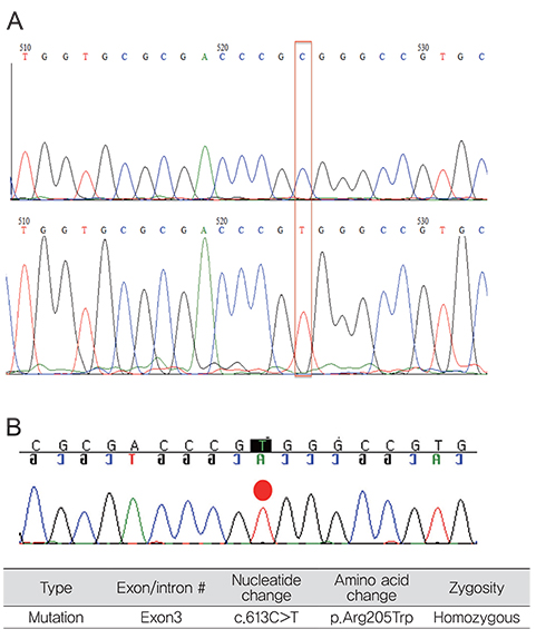
- To report a novel mutation within the CHST6 gene, as well as describe light and electron microscopic features of a case of macular corneal dystrophy. A 59-year old woman with macular corneal dystrophy in both eyes who had decreased visual acuity underwent penetrating keratoplasty. Further studies including light and electron microscopy, as well as DNA analysis were performed. Light microscopy of the cornea revealed glycosaminoglycan deposits in the keratocytes and endothelial cells, as well as extracellularly within the stroma. All samples stained positively with alcian blue, colloidal iron, and periodic acid-Schiff. Electron microscopy showed keratocytes distended by membrane-bound intracytoplasmic vacuoles containing electron-dense fibrillogranular material. These vacuoles were present in the endothelial cells and between stromal lamellae. Some of the vacuoles contained dense osmophilic whorls. A novel homozygous mutation (c.613 C>T [p.Arg205Trp]) was identified within the whole coding region of CHST6. A novel CHST6 mutation was detected in a Korean macular corneal dystrophy patient.
Keyword
MeSH Terms
-
Corneal Dystrophies, Hereditary/diagnosis/*genetics/metabolism
Corneal Keratocytes/ultrastructure
DNA/*genetics
DNA Mutational Analysis
Female
Humans
Microscopy, Electron
Middle Aged
*Mutation, Missense
Pedigree
Polymerase Chain Reaction
Republic of Korea
Sulfotransferases/*genetics/metabolism
DNA
Sulfotransferases
Figure
Reference
-
1. McTigue JW. The human cornea: a light and electron microscopic study of the normal cornea and its alterations in various dystrophies. Trans Am Ophthalmol Soc. 1967; 65:591–660.2. Donnenfeld ED, Cohen EJ, Ingraham HJ, et al. Corneal thinning in macular corneal dystrophy. Am J Ophthalmol. 1986; 101:112–113.3. Jee DH, Lee YD, Kim MS. Epidemiology of corneal dystrophy in Korea. J Korean Ophthalmol Soc. 2003; 44:581–587.4. Santo RM, Yamaguchi T, Kanai A, et al. Clinical and histopathologic features of corneal dystrophies in Japan. Ophthalmology. 1995; 102:557–567.5. Akama TO, Nishida K, Nakayama J, et al. Macular corneal dystrophy type I and type II are caused by distinct mutations in a new sulphotransferase gene. Nat Genet. 2000; 26:237–241.6. Dang X, Zhu Q, Wang L, et al. Macular corneal dystrophy in a Chinese family related with novel mutations of CHST6. Mol Vis. 2009; 15:700–705.7. Weiss JS, Moller HU, Lisch W, et al. The IC3D classification of the corneal dystrophies. Cornea. 2008; 27:Suppl 2. S1–S83.8. Snip RC, Kenyon KR, Green WR. Macular corneal dystrophy: ultrastructural pathology of corneal endothelium and Descemet's membrane. Invest Ophthalmol. 1973; 12:88–97.9. Klintworth GK, Vogel FS. Macular corneal dystrophy: an inherited acid mucopolysaccharide storage disease of the corneal fibroblast. Am J Pathol. 1964; 45:565–586.10. Hassell JR, Newsome DA, Krachmer JH, Rodrigues MM. Macular corneal dystrophy: failure to synthesize a mature keratan sulfate proteoglycan. Proc Natl Acad Sci U S A. 1980; 77:3705–3709.11. El-Ashry MF, Abd El-Aziz MM, Wilkins S, et al. Identification of novel mutations in the carbohydrate sulfotransferase gene (CHST6) causing macular corneal dystrophy. Invest Ophthalmol Vis Sci. 2002; 43:377–382.12. Musselmann K, Hassell JR. Focus on molecules: CHST6 (carbohydrate sulfotransferase 6; corneal N-acetylglucosamine-6-sulfotransferase). Exp Eye Res. 2006; 83:707–708.13. Aldave AJ, Yellore VS, Thonar EJ, et al. Novel mutations in the carbohydrate sulfotransferase gene (CHST6) in American patients with macular corneal dystrophy. Am J Ophthalmol. 2004; 137:465–473.14. Liu NP, Dew-Knight S, Rayner M, et al. Mutations in corneal carbohydrate sulfotransferase 6 gene (CHST6) cause macular corneal dystrophy in Iceland. Mol Vis. 2000; 6:261–264.15. Warren JF, Aldave AJ, Srinivasan M, et al. Novel mutations in the CHST6 gene associated with macular corneal dystrophy in southern India. Arch Ophthalmol. 2003; 121:1608–1612.
- Full Text Links
- Actions
-
Cited
- CITED
-
- Close
- Share
- Similar articles
-
- Cases of Macular Corneal Dystrophy with a Family History
- N102S Mutation of UBIAD1 Gene in a Family with Schnyder Crystalline Corneal Dystrophy
- Granular Corneal Dystrophy
- A Novel Decorin Gene Mutation in Congenital Hereditary Stromal Dystrophy: A Korean Family
- Four Cases of Avellino Corneal Dystrophy Concurrent with Floppy Eyelid Syndrome


