N102S Mutation of UBIAD1 Gene in a Family with Schnyder Crystalline Corneal Dystrophy
- Affiliations
-
- 1Corneal Dystrophy Research Institute, Department of Ophthalmology, Yonsei University College of Medicine, Seoul, Korea. eungkkim@yuhs.ac
Abstract
- PURPOSE
Schnyder crystalline corneal dystrophy (SCCD) is an autosomal dominant disease characterized by progressive central corneal opacification and premature development of peripheral arcus in the cornea. This disease results from a point mutation of UBIAD1 in chromosome 1p34-36. Until now, 15 different mutations of UBIAD1 gene on chromosome 1p34-36 have been reported for Schnyder crystalline corneal dystrophy. More point mutations are expected to be added to the list in the future. Schnyder crystalline corneal dystrophy is a rare disease, with only three reported cases in Korea, although there has been no report of a genetically confirmed case of the disease.
CASE SUMMARY
We encountered six patients with an N102S mutation of UBIAD1, who are from a family of two generation with 12 family members. Genetic confirmation for Schnyder crystalline corneal dystrophy was performed on these patients. This was the first report of a genetic confirmation of Schnyder crystalline corneal dystrophy in Korea. We will discuss our cases along with a review of the related literature.
MeSH Terms
Figure
-

Figure 1. Pedigree of the study family. Squares indicate male and circles indicate female. Slash means family member who was deceased, whereas blackened symbols represent the individuals who were affected by Schnyder crystalline corneal dystrophy. Black arrow indicates proband.

Figure 2. External photographs of the cornea of 38-year-old female(II:4, the proband) with BCVA of 0.8(OU) show bilateral deposition of anterior stromal crystals, corneal arcus and stromal haze. OD(A, B), OS(C, D).

Figure 3. Anterior segment Fourier domain OCT (A) and external photograph (B) of the right cornea of 38-year-old female (II:4) with BCVA of 0.8 show deposition of typical central subepithelial and anterior stromal crystals within anterior one third of the corneal depth. Crystalline deposits splitted into spindle shape (white arrow). Bowman layer was broken after crystalline deposits were dropped out (white wedge).
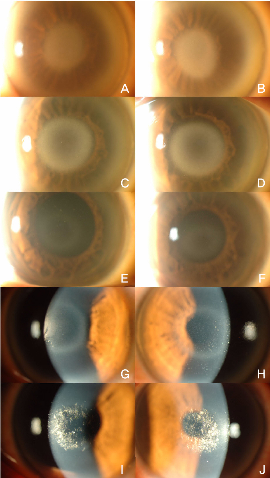
Figure 4. Slit-lamp photographs of affected patients' eyes (OD:A, C, E, G, I. OS:B, D, F, H, J.). (A, B) Both cornea of the 48-year-old female (II:2) shows with BCVA 0.4 (OU) show diffuse corneal opacifications and arcus lipoides. (C, D) Both cornea of the 44-year-old female (II: 3) shows with BCVA 0.6 (OU) show diffuse garland corneal opacifications and arcus lipoides. No crystals are apparent in the corneal stroma. (E, F) Both cornea of the 31-year-old female (II:7) shows show minimal garland stromal haze. (G, H) Both cornea of the 25-year-old female (III:1) shows with BCVA 0.8 (OU) show mild garland anterior stromal haze. (I, J) Both cornea of the 17-year-old male (III:3) shows with BCVA 0.8 (OD) 0.9 (OS) show dominant deposition of subepithelial crystals with slight stromal haze.

Figure 5. (A) Sequence chromatogram of the proband showing heterozygous mutation N102S in exon 1, forward reading. (B) Reverse reading of the same mutation, showing N102S.
Reference
-
References
1. Weiss JS. Visual morbidity in thirty-four families with Schnyder crystalline corneal dystrophy (an American Ophthalmological Society thesis). Trans Am Ophthalmol Soc. 2007; 105:616–48.2. Shearman AM, Hudson TJ, Andresen JM, et al. The gene for schnyder's crystalline corneal dystrophy maps to human abdominal 1p34.1-p36. Hum Mol Genet. 1996; 5:1667–72.3. Aldave AJ, Rayner SA, Principe AH, et al. Analysis of fifteen abdominal candidate genes for Schnyder crystalline corneal dystrophy. Mol Vis. 2005; 11:713–6.4. Riebeling P, Polz S, Tost F, et al. Schnyder's crystalline corneal dystrophy. Further narrowing of the linkage interval at abdominal 1p34.1-p36? Ophthalmologe. 2003; 100:979–83.5. Theendakara V, Tromp G, Kuivaniemi H, et al. Fine mapping of the Schnyder's crystalline corneal dystrophy locus. Hum Genet. 2004; 114:594–600.
Article6. Weiss JS, Kruth HS, Kuivaniemi H, et al. Mutations in the UBIAD1 gene on chromosome short arm 1, region 36, cause Schnyder crystalline corneal dystrophy. Invest Ophthalmol Vis Sci. 2007; 48:5007–12.7. Kim HB, Kong YT, Hong YJ. Two Cases of Hereditary Crystaline Corneal Dystrophy of Schnyder. J Korean Ophthalmol Soc. 1973; 14:387–91.8. Kim JH, Myong YW. Histopathological Findings of Schnyder's Crystalline Corneal Dystrophy. J Korean Ophthalmol Soc. 1995; 36:1363–9.9. Jung SC, Myong YW. A Case of Spontaneous Regression of Schnyder's Crystalline Corneal Dystrophy. J Korean Ophthalmol Soc. 2000; 41:1441–4.10. Orr A, Dube MP, Marcadier J, et al. Mutations in the UBIAD1 gene, encoding a potential prenyltransferase, are causal for Schnyder crystalline corneal dystrophy. PLoS One. 2007; 2:e685.
Article11. Kobayashi A, Fujiki K, Murakami A, Sugiyama K. In vivo laser confocal microscopy findings and mutational analysis for Schnyder's crystalline corneal dystrophy. Ophthalmology. 2009; 116:1029–37.
Article12. Weiss JS, Kruth HS, Kuivaniemi H, et al. Genetic analysis of 14 families with Schnyder crystalline corneal dystrophy reveals clues to UBIAD1 protein function. Am J Med Genet A. 2008; 146:271–83.
Article13. Jing Y, Liu C, Xu J, Wang L. A novel UBIAD1 mutation abdominal in a Chinese family with Schnyder crystalline corneal dystrophy. Mol Vis. 2009; 15:1463–9.14. Weiss JS. Schnyder crystalline dystrophy sine crystals. Recommendation for a revision of nomenclature. Ophthalmology. 1996; 103:465–73.15. van Went JM WF. En zeldzame erfelijke hoornvliessandoening. Niederl Tijdschr Geneesks. 1924; 68:2996–7.16. WF S. Mitteilung über einen neuen Typus von familiärer Hornhauterkrankung. Schweiz Med Wochenschr. 1929; 10:559–71.17. Vesaluoma MH, Linna TU, Sankila EM, et al. In vivo confocal abdominal of a family with Schnyder crystalline corneal dystrophy. Ophthalmology. 1999; 106:944–51.18. Weller RO, Rodger FC. Crystalline stromal dystrophy: histochemistry and ultrastructure of the cornea. Br J Ophthalmol. 1980; 64:46–52.
Article19. Delleman JW, Winkelman JE. Degeneratio corneae cristallinea hereditaria. A clinical, genetical and histological study. Ophthalmologica. 1968; 155:409–26.20. Herring JH, Phillips D, McCaa CS. Phototherapeutic abdominal for Schnyder's central crystalline dystrophy. J Refract Surg. 1999; 15:4891989; 8:135–40.21. Bron AJ. Corneal changes in the dislipoproteinaemias. Cornea. 1989; 8:135–40.
Article22. Brownstein S, Jackson WB, Onerheim RM. Schnyder's abdominal corneal dystrophy in association with hyperlipoproteinemia: histopathological and ultrastructural findings. Can J Ophthalmol. 1991; 26:273–9.23. Lisch W, Weidle EG, Lisch C, et al. Schnyder's dystrophy. Progression and metabolism. Ophthalmic Paediatr Genet. 1986; 7:45–56.
Article
- Full Text Links
-
- Actions
-
Cited
- CITED
-
- Close
- Share
-
- Similar articles
-
- A Case of Spontaneous Regression of Schnyder's Crystalline Corneal Dystrophy
- Histopathological Findings of Schnyder's Crystalline Corneal Dystrophy
- Two Cases of Hereditary Crystalline Corneal Dystrophy of Schnyder
- A case of Excimer Laser PRK in Schnyder's Dystrophy
- Cases of Macular Corneal Dystrophy with a Family History
