Correlation between skeletal and dental changes after mandibular setback surgery-first orthodontic treatment: Cone-beam computed tomography-generated half-cephalograms
- Affiliations
-
- 1Private Practice, Busan, Korea.
- 2Department of Orthodontics, Biomedical Research Institute, Pusan National University Hospital, Busan, Korea. kimyongil@pusan.ac.kr
- 3Department of Orthodontics, Dental Research Institute, Pusan National University Dental Hospital, Yangsan, Korea.
- KMID: 1787606
- DOI: http://doi.org/10.4041/kjod.2015.45.2.59
Abstract
OBJECTIVE
To investigate skeletal and dental changes after application of a mandibular setback surgery-first orthodontic treatment approach in cases of skeletal Class III malocclusion.
METHODS
A retrospective study of 34 patients (23 men, 11 women; mean age, 26.2 +/- 6.6 years) with skeletal Class III deformities, who underwent surgery-first orthodontic treatment, was conducted. Skeletal landmarks in the maxilla and mandible at three time points, pre-treatment (T0), immediate-postoperative (T1), and post-treatment (T2), were analyzed using cone-beam computed tomography (CBCT)-generated half-cephalograms.
RESULTS
The significant T0 to T1 mandibular changes occurred -9.24 +/- 3.97 mm horizontally. From T1 to T2, the mandible tended to move forward 1.22 +/- 2.02 mm, while the condylar position (Cd to Po-perpendicular plane) shifted backward, and the coronoid process (Cp to FH plane) moved vertically. Between T1 and T2, the vertical dimension changed significantly (p < 0.05). Changes in the vertical dimension were significantly correlated to T1 to T2 changes in the Cd to Po-perpendicular plane (r = -0.671, p = 0.034), and in the Cp to FH plane (r = 0.733, p = 0.016), as well as to T0 to T1 changes in the Cp to Po-perpendicular plane (r = 0.758, p = 0.011).
CONCLUSIONS
Greater alterations in the vertical dimension caused larger post-treatment (T2) stage skeletal changes. Studying the mandibular position in relation to the post-surgical vertical dimension emphasized the integral importance of vertical dimension control and proximal segment management to the success of surgery-first orthodontic treatment.
MeSH Terms
Figure
-
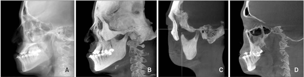
Figure 1 Generation of cone-beam computed tomography generated half-cephalograms. Raycast images. A, Maximum intensity projection (MIP) image. B, Coronid process and condyle MIP image. C, Maxillary and mandibular molar MIP image. D, Maxillary and mandibular molar MIP image.
Cited by 4 articles
-
Evaluation of stability after pre-orthodontic orthognathic surgery using cone-beam computed tomography: A comparison with conventional treatment
Hye-Rim Ann, Young-Soo Jung, Kee-Joon Lee, Hyoung-Seon Baik
Korean J Orthod. 2016;46(5):301-309. doi: 10.4041/kjod.2016.46.5.301.Comparison of postoperative changes in the distal and proximal segments between conventional and sliding mini-plate fixation following mandibular setback
Seong-Sik Kim, Kyoung-Ho Kwak, Ching-Chang Ko, Soo-Byung Park, Woo-Sung Son, Yong-Il Kim
Korean J Orthod. 2016;46(6):372-378. doi: 10.4041/kjod.2016.46.6.372.Comparison of changes in the transverse dental axis between patients with skeletal Class III malocclusion and facial asymmetry treated by orthognathic surgery with and without presurgical orthodontic treatment
Han-Sol Song, Sung-Hwan Choi, Jung-Yul Cha, Kee-Joon Lee, Hyung-Seog Yu
Korean J Orthod. 2017;47(4):256-267. doi: 10.4041/kjod.2017.47.4.256.The genial tubercle: A prospective novel landmark for the diagnosis of mandibular asymmetry
Seung-Youp Lee, Dong-Soon Choi, Insan Jang, Geun-Su Song, Bong-Kuen Cha
Korean J Orthod. 2017;47(1):50-58. doi: 10.4041/kjod.2017.47.1.50.
Reference
-
1. Park KR, Kim SY, Park HS, Jung YS. Surgery-first approach on patients with temporomandibular joint disease by intraoral vertical ramus osteotomy. Oral Surg Oral Med Oral Pathol Oral Radiol. 2013; 116:e429–e436.
Article2. Hernández-Alfaro F, Guijarro-Martínez R, Molina-Coral A, Badía-Escriche C. "Surgery first" in bimaxillary orthognathic surgery. J Oral Maxillofac Surg. 2011; 69:e201–e207.
Article3. Nagasaka H, Sugawara J, Kawamura H, Nanda R. "Surgery first" skeletal Class III correction using the Skeletal Anchorage System. J Clin Orthod. 2009; 43:97–105.4. Liou EJ, Chen PH, Wang YC, Yu CC, Huang CS, Chen YR. Surgery-first accelerated orthognathic surgery: postoperative rapid orthodontic tooth movement. J Oral Maxillofac Surg. 2011; 69:781–785.
Article5. Baek SH, Ahn HW, Kwon YH, Choi JY. Surgery-first approach in skeletal class III malocclusion treated with 2-jaw surgery: evaluation of surgical movement and postoperative orthodontic treatment. J Craniofac Surg. 2010; 21:332–338.
Article6. Liou EJ, Chen PH, Wang YC, Yu CC, Huang CS, Chen YR. Surgery-first accelerated orthognathic surgery: orthodontic guidelines and setup for model surgery. J Oral Maxillofac Surg. 2011; 69:771–780.
Article7. Hwang DS, Kim YI, Lee KM. Effect of intended manual condylar positioning on skeletal and dental changes in Skeletal Class III deformities: CBCT-generated half-cephalograms. J Craniomaxillofac Surg. 2014; 42:7–12.
Article8. Kim SJ, Park SB, Kim YI, Cho BH, Hwang DS. The reliability of cone-beam computed tomography (CBCT) - generated frontal cephalograms. J Craniomaxillofac Surg. 2012; 40:e331–e336.
Article9. Hwang HS, Lee KM, Uhm GS, Cho JH, McNamara JA Jr. Use of Reference Ear Plug to improve accuracy of lateral cephalograms generated from cone-beam computed tomography scans. Korean J Orthod. 2013; 43:54–61.
Article10. Lee JH, Kim MJ, Kim SM, Kwon OH, Kim YK. The 3D CT superimposition method using image fusion based on the maximum mutual information algorithm for the assessment of oral and maxillofacial surgery treatment results. Oral Surg Oral Med Oral Pathol Oral Radiol. 2012; 114:167–174.
Article11. Kang SH, Kim MK, Park SY, Lee JY, Park W, Lee SH. Early orthognathic surgery with three-dimensional image simulation during presurgical orthodontics in adults. J Craniofac Surg. 2011; 22:473–481.
Article12. Jeon J, Kim Y, Kim J, Kang H, Ji H, Son W. New bimaxillary orthognathic surgery planning and model surgery based on the concept of six degrees of freedom. Korean J Orthod. 2013; 43:42–52.
Article13. Hwang DS, Kim YI, Lee JY, Lee ST, Kim TH, Lee JM, et al. Evaluation of skeletal stability following twojaw surgery via surgery first orthodontic treatment in class iii malocclusion. J Korean Assoc Maxillofac Plast Reconstr Surg. 2011; 33:407–412.14. Kim YI, Park SB, Jung YH, Hwang DS, Lee JY. Evaluation of intersegmental displacement according to osteosynthesis method for mandibular setback sagittal split ramus osteotomy using cone-beam computed tomographic superimposition. J Oral Maxillofac Surg. 2012; 70:2893–2898.
Article
- Full Text Links
-
- Actions
-
Cited
- CITED
-
- Close
- Share
-
- Similar articles
-
- Immediate changes in the mandibular dentition after maxillary molar distalization using headgear
- Qualitative correlation between postoperatively increased vertical dimension and mandibular position in skeletal class III using partial-least-square path modeling
- Use of Head Posture Aligner to improve accuracy of frontal cephalograms generated from cone-beam CT scans
- Three dimensional cone-beam CT study of upper airway change after mandibular setback surgery for skeletal Class III malocclusion patients
- Use of Reference Ear Plug to improve accuracy of lateral cephalograms generated from cone-beam computed tomography scans
