Cyclin-Dependent Kinase Inhibitor p27(Kip1) Controls Growth and Cell Cycle Progression in Human Uterine Leiomyoma
- Affiliations
-
- 1Department of Obstetrics and Gynecology, Keimyung University, School of Medicine, Daegu, Korea. c0035@dsmc.or.kr
- 2Department of Pathology, Keimyung University, School of Medicine, Daegu, Korea.
- 3Department of Oncology, Lombardi Cancer Center, Georgetown University, USA.
- KMID: 1785827
- DOI: http://doi.org/10.3346/jkms.2008.23.4.667
Abstract
- The molecular mechanism of the cell-cycle machinery in uterine leiomyoma has not yet been fully elucidated. Among the various types of cell-cycle regulators, p27(Kip1)(p27) is considered to be a potent tumor suppressor. To provide further molecular basis for understanding the progression of uterine leiomyoma, our objective was to evaluate the expression level of p27 in normal myometrium and uterine leiomyoma tissue and its effect on cytogenic growth. Western blot analysis, real-time polymerase chain reaction (PCR) and immunohistochemical staining revealed that p27 protein and messenger RNA were down-regulated in uterine leiomyoma tissue and cultured cells compared to normal myometerium. Full-length human p27 cDNA was transferred using a replication-deficient recombinant adenoviral vector (Ad.p27) into uterine leiomyoma cells and evaluated the effect on cell proliferation. Transfection of Ad.p27 into uterine leiomyoma cells resulted in the induction of apoptosis, reduction in viability and proliferation of uterine leiomyoma cells. Our results suggest a new paradigm that down-regulated p27 protein expression is the possible underlying mechanism for the growth of uterine leiomyoma and over-expression of p27 induces cell death. This study provides better understanding of the control exerted by p27 in regulating growth and disease progression of uterine leiomyoma.
MeSH Terms
Figure
-
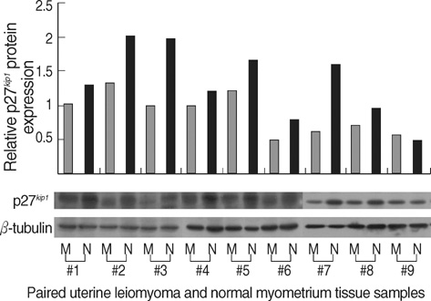
Fig. 1 Decreased p27 protein expression levels in human uterine leiomyoma tissue.Total protein was isolated from uterine leiomyoma and normal myometrial tissues. Nine pairs of tumor and normal tissues; i.e., each pair of samples was obtained from same patient - M; uterine leiomyoma, N; normal myometrium, 5 of the tissue samples belonged to the proliferative phase (#1 to #5) and the rest to the secretory phase (#6 to #9). Fifty micrograms of the total isolated protein were resolved by SDS-polyacrylamide gel electrophoresis and electroblotted onto PVDF membranes. The blot was incubated with antibody against p27. Reactive bands were visualized with an ECL labeling and detection system. β-tubulin was used as the loading control. Densitometric analysis was also done to the western blots to determine the relative protein expression levels.
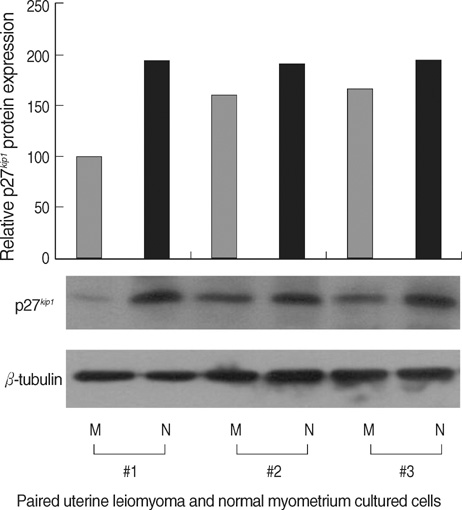
Fig. 2 Decreased p27 protein expression levels in cultured human uterine leiomyoma tissue. Total protein was isolated from cultured, well-grown, confluent uterine leiomyoma and normal myometrium tissue samples (3 pairs of tumor and normal tissue, obtained from same patient - M; uterine leiomyoma, N; normal myometrium, 2 of the cultured cells were from proliferative phase tissue samples (#1 and #2) and #3 was cultured cells of the secretory phase tissue sample). Fifty micrograms of the total isolated protein were resolved by SDS-polyacrylamide gel electrophoresis and electroblotted onto PVDF membranes. The blot was incubated with antibody against p27. Reactive bands were visualized with an ECL labeling and detection system. β-tubulin was used as the loading control. Densitometric analysis was also done to the Western blots to determine the relative protein expression levels.
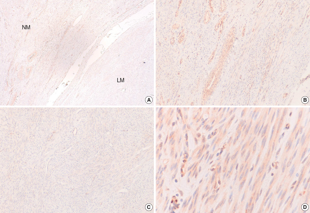
Fig. 3 Comparison of immunohistochemical staining for p27 protein. Immunohistochemical staining for p27 formalin-fixed paraffin-embedded sections of uterine leiomyoma (LM) and myometrial tissues (NM). (A) Myometrial smooth muscle cells showed more predominant immunostaining for p27 protein than the leiomyoma cells (magnification ×40), (B). Normal myometrial cells showed positive for p27 protein (magnification ×100), (C). Leiomyoma cells showed negative for p27 protein (magnification ×100), (D). Cytoplasmic and nuclear brown-staining cells are positive for p27 protein expression (magnification ×400).
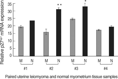
Fig. 4 Decreased p27 mRNA levels in human uterine leiomyoma tissue. Total RNA was isolated from uterine leiomyoma and normal myometrial tissues (4 pairs of tumor and normal tissues; i.e., each pair of samples was obtained from same patient; tissue samples #1 and #2 were isolated at the proliferative phase and tissue samples #3 and #4 were isolated at the secretory phases of the menstrual cycle). cDNA templates for RT were prepared from the total RNA extracted. The level of p27 mRNA was determined by quantitative real-time PCR. Relative p27 mRNA levels (normalized to GAPDH) in paired samples represented as M; uterine leiomyoma, N; normal myometrium. *, p<0.05; **, p<0.01.

Fig. 5 Effect of Ad.p27 transfection on uterine leiomyoma cells. Cultured uterine leiomyoma cells were transfected with Ad.null and Ad.p27 recombinant, non-replicating adenoviral vectors. Twenty-four hours after transfection, immunoblotting was performed to detect the expression levels of p27, p21, Rb, cdk2 and cdk4. β-tubulin was used as the loading control. Transfection of Ad.p27 onto uterine leiomyoma cells caused increased expression levels of p27 and dephosphorylation of Rb.
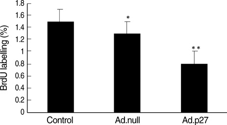
Fig. 6 Inhibition of the synthesis of DNA in uterine leiomyoma cells by Ad.p27. The antiproliferative effects were determined after transfecting uterine leiomyoma cells for 24 hr with Ad.null and Ad.p27 adenoviral vectors and labeling with BrdU (10 µM) for 24 hr. Results are expressed as percentage inhibition of BrdU incorporation relative to control cells. Values are the means (±SD) of three experiments with triplicate determinations. *, p<0.05; **, p<0.01.
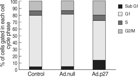
Fig. 7 Effect of Ad.p27 on the cell cycle profile. Ad.null and Ad.p27 transfected uterine leiomyoma cells were harvested, fixed, stained with PI, and analyzed by flow cytometry analysis. The values represent the number of cells in each phase of the cell cycle as a percentage (%) of total cells. Following transfection with Ad.p27, the growth of uterine leiomyoma cells was affected with an increase in the percentage of cells in the sub-G1 phase.
Reference
-
1. Buttram VC, Reiter RC. Uterine leiomyomata: etiology, symptomatology, and management. Fertil Steril. 1981. 36:433–445.2. Sherr CJ, Roberts JM. CDK inhibitors: positive and negative regulators of G1-phase progression. Genes Dev. 1999. 13:1501–1512.
Article3. Tchernev G, Orfanos CE. Downregulation of cell cycle modulators p21, p27, p53, Rb and proapoptotic Bcl-2-related proteins Bax and Bak in cutaneous melanoma is associated with worse patient prognosis: preliminary findings. J Cutan Pathol. 2007. 34:247–256.
Article4. Matsuda Y, Ichida T. p16 and p27 are functionally correlated during the progress of hepatocarcinogenesis. Med Mol Morphol. 2006. 39:169–175.
Article5. Shariat SF, Ashfaq R, Sagalowsky AI, Lotan Y. Predictive value of cell cycle biomarkers in nonmuscle invasive bladder transitional cell carcinoma. J Urol. 2007. 177:481–487.
Article6. Saltman B, Singh B, Hedvat CV, Wreesmann VB, Ghossein R. Patterns of expression of cell cycle/apoptosis genes along the spectrum of thyroid carcinoma progression. Surgery. 2006. 140:899–905.
Article7. Tan P, Cady B, Wanner M, Worland P, Cukor B, Magi-Galluzzi C, Lavin P, Draetta G, Pagano M, Loda M. The cell cycle inhibitor p27 is an independent prognostic marker in small (T1a,b) invasive breast carcinomas. Cancer Res. 1997. 57:1259–1263.8. Tsihlias J, Kapusta LR, DeBoer G, Morava-Protzner I, Zbieranowski I, Bhattacharya N, Catzavelos GC, Klotz LH, Slingerland JM. Loss of cyclin-dependent kinase inhibitor p27Kip1 is a novel prognostic factor in localized human prostate adenocarcinoma. Cancer Res. 1998. 58:542–548.9. Lahav-Baratz S, Ben-Izhak O, Sabo E, Ben-Eliezer S, Lavie O, Ishai D, Ciechanover A, Dirnfeld M. Decreased level of the cell cycle regulator p27 and increased level of its ubiquitin ligase Skp2 in endometrial carcinoma but not in normal secretory or in hyperstimulated endometrium. Mol Hum Reprod. 2004. 10:567–572.
Article10. Tong W, Pollard JW. Progesterone inhibits estrogen-induced cyclin D1 and cdk4 nuclear translocation, cyclin E- and cyclin A-cdk2 kinase activation, and cell proliferation in uterine epithelial cells in mice. Mol Cell Biol. 1999. 19:2251–2264.
Article11. Shiozawa T, Horiuchi A, Kato K, Obinata M, Konishi I, Fujii S, Nikaido T. Up-regulation of p27Kip1 by progestins is involved in the growth suppression of the normal and malignant human endometrial glandular cells. Endocrinology. 2001. 142:4182–4188.
Article12. Slingerland J, Pagano M. Regulation of the cdk inhibitor p27 and its deregulation in cancer. J Cell Physiol. 2000. 183:10–17.
Article13. Dobashi Y, Noguchi T, Nasuno S, Katayama K, Kameya T. CDK-inhibitors-associated kinase activity: a possible determinant of malignant potential in smooth muscle tumors of the external soft tissue. Int J Cancer. 2001. 94:353–362.
Article14. Leiser AL, Anderson SE, Nonaka D, Chuai S, Olshen AB, Chi DS, Soslow RA. Apoptotic and cell cycle regulatory markers in uterine leiomyosarcoma. Gynecol Oncol. 2006. 101:86–91.
Article15. Luo X, Ding L, Xu J, Chegini N. Gene expression profiling of leiomyoma and myometrial smooth muscle cells in response to transforming growth factor-beta. Endocrinology. 2005. 146:1097–1118.16. Liu S, Bishop WR, Liu M. Differential effects of cell cycle regulatory protein p21 (WAF1/Cip1) on apoptosis and sensitivity to cancer chemotherapy. Drug Resist Updat. 2003. 6:183–195.17. Park KH, Seol JY, Yoo CG, Kim YW, Han SK, Lee EH, Kim CM, Shim YS, Lee CT. Adenovirus expressing p27Kip1 induces growth arrest of lung cancer cell lines and suppresses the growth of established lung cancer xenografts. Lung Cancer. 2001. 31:149–155.18. Sherr CJ, Roberts JM. Inhibitors of mammalian G1 cyclin-dependent kinases. Genes Dev. 1995. 9:1149–1163.19. Fredersdorf S, Burns J, Milne AM, Packham G, Fallis L, Gillett CE, Royds JA, Peston D, Hall PA, Hanby AM, Barnes DM, Shousha S, O'Hare MJ, Lu X. High level expression of p27Kip1 and cyclin D1 in some human breast cancer cells: inverse correlation between the expression of p27Kip1 and degree of malignancy in human breast and colorectal cancers. Proc Natl Acad Sci USA. 1997. 94:6380–6385.
Article20. Ciaparrone M, Yamamoto H, Yao Y, Sgambato A, Cattoretti G, Tomita N, Monden T, Rotterdam H, Weinstein IB. Localization and expression of p27Kip1 in multistage colorectal carcinogenesis. Cancer Res. 1998. 58:114–122.21. Singh SP, Lipman J, Goldman H, Ellis FH Jr, Aizenman L, Cangi MG, Signoretti S, Chiaur DS, Pagano M, Loda M. Loss or altered subcellular localization of p27 in Barrett's associated adenocarcinoma. Cancer Res. 1998. 58:1730–1735.22. Chen TP, Chen CM, Chang HW, Wang JS, Chang WC, Hsu SI, Cho CL. Increased expression of SKP2 and phospho-MAPK/ERK1/2 and decreased expression of p27 during tumor progression of cervical neoplasms. Gynecol Oncol. 2007. 104:516–523.
Article23. Pateras IS, Apostolopoulou K, Koutsami M, Evangelou K, Tsantoulis P, Liloglou T, Nikolaidis G, Sigala F, Kittas C, Field JK, Kotsinas A, Gorgoulis VG. Downregulation of the KIP family members p27 (KIP1) and p57 (KIP2) by SKP2 and the role of methylation in p57 (KIP2) inactivation in nonsmall cell lung cancer. Int J Cancer. 2006. 119:2546–2556.24. Dvorackova J, Uvirova M. A molecularly genetic determination of prognostic factors of the prostate cancer and their relationships to expression of protein p27kip1. Neoplasma. 2007. 54:149–154.25. Akiyama T, Ohuchi T, Sumida S, Matsumoto K, Toyoshima K. Phosphorylation of the retinoblastoma protein by cdk2. Proc Natl Acad Sci USA. 1992. 89:7900–7904.
Article26. Weinberg RA. Tumor suppressor genes. Science. 1991. 254:1138–1146.
Article27. Hong FD, Chen J, Donovan S, Schneider N, Nisen PD. Taxol, vincristine or nocodazole induces lethality in G1-checkpoint-defective human astrocytoma U373MG cells by triggering hyperploid progression. Carcinogenesis. 1999. 20:1161–1168.
Article
- Full Text Links
-
- Actions
-
Cited
- CITED
-
- Close
- Share
-
- Similar articles
-
- MicroRNA-186 targets SKP2 to induce p27(Kip1)-mediated pituitary tumor cell cycle deregulation and modulate cell proliferation
- The Relevance of Women's Diseases, Jun Activation-domain Binding Protein 1 (JAB1) and p27(kip1)
- The function of p27(KIP1) during tumor development
- Prognostic Implications of Cyclin B1, p34cdc2, p27(Kip1) and p53 Expression in Gastric Cancer
- p27(Kip1) and Ki-67 Expression in Mucoepidermoid Carcinoma
