Diagnostic Value of 64-Slice Dual-Source CT Coronary Angiography in Patients with Atrial Fibrillation: Comparison with Invasive Coronary Angiography
- Affiliations
-
- 1Department of Radiology, Zhejiang Hospital, Zhejiang Province, 310013, China. zhangyp31113@163.com
- KMID: 1783207
- DOI: http://doi.org/10.3348/kjr.2011.12.4.416
Abstract
OBJECTIVE
We wanted to evaluate the image quality and diagnostic value of 64-slice dual-source computed tomography (DSCT) coronary angiography in patients with atrial fibrillation (Afib).
MATERIALS AND METHODS
The coronary arteries of 22 Afib patients seen on DSCT were classified into 15 segments and the imaging quality (excellent, good, moderate and poor) and significant stenoses (> or = 50%) were evaluated by two radiologists who were blinded to the conventional coronary angiography (CAG) results. The sensitivity, specificity, positive predictive value (PPV) and negative predictive value (NPV) for detecting important coronary artery stenosis were calculated. McNemar test was used to determine any significant difference between DSCT and CAG, and Cohen's Kappa statistics were calculated for the intermodality and interobserver agreement.
RESULTS
The mean heart rate was 89 +/- 8.3 bpm (range: 80-118 bpm). A range from 250 msec to 300 msec within the RR interval was the optimal reconstruction interval for the patients with Afib. The respective overall sensitivity, specificity, PPV and NPV values were 74%, 97%, 81% and 96% for reader 1 and 72%, 98%, 85% and 96% for reader 2. No significant difference between DSCT and CAG was found for detecting a significant stenosis (reader 1, p = 1.0; reader 2, p = 0.727). Cohen's Kappa statistics demonstrated good intermodality and interobserver agreement.
CONCLUSION
64-slice DSCT coronary angiography provides good image quality in patients with atrial fibrillation without the need for controlling the heart rate. DSCT can be used for ruling out significant stenosis in patients with atrial fibrillation with its high NPV for detecting in important stenosis.
Keyword
MeSH Terms
-
Aged
Aged, 80 and over
Algorithms
Atrial Fibrillation/*radiography
Contrast Media/diagnostic use
Coronary Angiography/*methods
Coronary Disease/*radiography
Echocardiography
Electrocardiography
Female
Heart Rate
Humans
Iohexol/analogs & derivatives/diagnostic use
Male
Middle Aged
Prospective Studies
Radiation Dosage
Radiographic Image Interpretation, Computer-Assisted
Tomography, X-Ray Computed/*methods
Figure
-

Fig. 1 CT angiography of 86-year-old man with atrial fibrillation (mean heart rate: 93 bpm, range: 66-143 bpm). A, B. Curved multiplanar image (A) and maximum intensity projection image (B) showing two slight stenoses in right coronary artery segments 1 and 2 (arrows). C. Invasive angiography confirms these two lesions (arrows). D. Curved multiplanar image showing moderate stenosis and accompanying atherosclerotic lesions in left anterior descending artery segments 6 and 7 (arrows). E. Invasive angiography of left anterior descending artery confirms lesions (arrows). F. Echocardiography is shown (reconstruction at 250 ms of cardiac cycle).
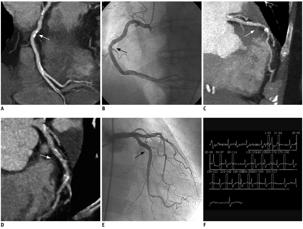
Fig. 2 CT angiography of 70-year-old woman with atrial fibrillation (mean heart rate: 87 bpm, range: 60-125 bpm). A. Curved multiplanar image showing slight stenosis in right coronary artery segment 1 (arrow). B. Invasive angiography of right coronary artery confirms presence (arrow) of mild lesion. C, D. Maximum intensity projection image (C) and curved multiplanar image (D) showing slight stenosis and accompanying atherosclerotic lesions in left anterior descending artery segment 7 (arrows). E. Invasive angiography confirms presence of lesion (arrow). F. Echocardiography is shown (reconstruction at 300 ms of cardiac cycle).

Fig. 3 Relationship between heart rate and cardiac phase that provided for optimal image quality.
Reference
-
1. Schoepf UJ, Becker CR, Ohnesorge BM, Yucel EK. CT of coronary artery disease. Radiology. 2004. 232:18–37.2. Schoenhagen P, Halliburton SS, Stillman AE, Kuzmiak SA, Nissen SE, Tuzcu EM, et al. Noninvasive imaging of coronary arteries: current and future role of multi-detector row CT. Radiology. 2004. 232:7–17.3. Raff GL, Gallagher MJ, O'Neill WW, Goldstein JA. Diagnostic accuracy of noninvasive coronary angiography using 64-slice spiral computed tomography. J Am Coll Cardiol. 2005. 46:552–557.4. Sun Z, Jiang W. Diagnostic value of multislice computed tomography angiography in coronary artery disease: a meta-analysis. Eur J Radiol. 2006. 60:279–286.5. Heuschmid M, Kuettner A, Schroeder S, Trabold T, Feyer A, Seemann MD, et al. ECG-gated 16-MDCT of the coronary arteries: assessment of image quality and accuracy in detecting stenoses. AJR Am J Roentgenol. 2005. 184:1413–1419.6. Pugliese F, Mollet NR, Runza G, van Mieghem C, Meijboom WB, Malagutti P, et al. Diagnostic accuracy of non-invasive 64-slice CT coronary angiography in patients with stable angina pectoris. Eur Radiol. 2006. 16:575–582.7. Ha EJ, Kim Y, Cheung JY, Shim SS. Coronary artery disease in asymptomatic young adults: its prevalence according to coronary artery disease risk stratification and the CT characteristics. Korean J Radiol. 2010. 11:416–432.8. Flohr TG, McCollough CH, Bruder H, Petersilka M, Gruber K, Suss C, et al. First performance evaluation of a dual-source CT (DSCT) system. Eur Radiol. 2006. 16:256–268.9. Achenbach S, Ropers D, Kuettner A, Flohr T, Ohnesorge B, Bruder H, et al. Contrast-enhanced coronary artery visualization by dual-source computed tomography--initial experience. Eur J Radiol. 2006. 57:331–335.10. Johnson TR, Nikolaou K, Wintersperger BJ, Leber AW, von Ziegler F, Rist C, et al. Dual-source CT cardiac imaging: initial experience. Eur Radiol. 2006. 16:1409–1415.11. Austen WG, Edwards JE, Frye RL, Gensini GG, Gott VL, Griffith LS, et al. A reporting system on patients evaluated for coronary artery disease. Report of the Ad Hoc Committee for Grading of Coronary Artery Disease, Council on Cardiovascular Surgery, American Heart Association. Circulation. 1975. 51:5–40.12. Johnson TR, Nikolaou K, Busch S, Leber AW, Becker A, Wintersperger BJ, et al. Diagnostic accuracy of dual-source computed tomography in the diagnosis of coronary artery disease. Invest Radiol. 2007. 42:684–691.13. Althen JN. Automatic tube-current modulation in CT--a comparison between different solutions. Radiat Prot Dosimetry. 2005. 114:308–312.14. Pasricha SS, Nandurkar D, Seneviratne SK, Cameron JD, Crossett M, Schneider-Kolsky ME, et al. Image quality of coronary 320-MDCT in patients with atrial fibrillation: initial experience. AJR Am J Roentgenol. 2009. 193:1514–1521.15. Oncel D, Oncel G, Tastan A. Effectiveness of dual-source CT coronary angiography for the evaluation of coronary artery disease in patients with atrial fibrillation: initial experience. Radiology. 2007. 245:703–711.16. Rist C, Johnson TR, Muller-Starck J, Arnoldi E, Saam T, Becker A, et al. Noninvasive coronary angiography using dual-source computed tomography in patients with atrial fibrillation. Invest Radiol. 2009. 44:159–167.17. Marwan M, Pflederer T, Schepis T, Lang A, Muschiol G, Ropers D, et al. Accuracy of dual-source computed tomography to identify significant coronary artery disease in patients with atrial fibrillation: comparison with coronary angiography. Eur Heart J. 2010. 31:2230–2237.18. Leschka S, Scheffel H, Desbiolles L, Plass A, Gaemperli O, Valenta I, et al. Image quality and reconstruction intervals of dual-source CT coronary angiography: recommendations for ECG-pulsing windowing. Invest Radiol. 2007. 42:543–549.19. Stein PD, Beemath A, Kayali F, Skaf E, Sanchez J, Olson RE. Multidetector computed tomography for the diagnosis of coronary artery disease: a systematic review. Am J Med. 2006. 119:203–216.20. Herzog C, Arning-Erb M, Zangos S, Eichler K, Hammerstingl R, Dogan S, et al. Multi-detector row CT coronary angiography: influence of reconstruction technique and heart rate on image quality. Radiology. 2006. 238:75–86.21. Leschka S, Wildermuth S, Boehm T, Desbiolles L, Husmann L, Plass A, et al. Noninvasive coronary angiography with 64-section CT: effect of average heart rate and heart rate variability on image quality. Radiology. 2006. 241:378–385.22. Scheffel H, Alkadhi H, Plass A, Vachenauer R, Desbiolles L, Gaemperli O, et al. Accuracy of dual-source CT coronary angiography: First experience in a high pre-test probability population without heart rate control. Eur Radiol. 2006. 16:2739–2747.23. Wang Y, Zhang Z, Kong L, Song L, Merges RD, Chen J, et al. Dual-source CT coronary angiography in patients with atrial fibrillation: comparison with single-source CT. Eur J Radiol. 2008. 68:434–441.24. Rixe J, Rolf A, Conradi G, Elsaesser A, Moellmann H, Nef HM, et al. Image quality on dual-source computed-tomographic coronary angiography. Eur Radiol. 2008. 18:1857–1862.
- Full Text Links
-
- Actions
-
Cited
- CITED
-
- Close
- Share
-
- Similar articles
-
- Diagnostic accuracy of 64-slice multi-detector CT coronary angiography in the evaluation of coronary artery disease
- Noninvasive Detection of Coronary Atherosclerotic Plaques and Assessment of Stenosis Degree at Multidetector CT Coronary Angiography
- Diagnostic accuracy of 64-slice multidetector CT coronary angiography for the evaluation of coronary artery disease
- The Diagnostic Accuracy of the 64-slice Multi-detector CT Coronary Angiography for the Assessment of Coronary Artery Stenosis in Symptomatic Patients
- Giant coronary aneurysm caused by Kawasaki disease: consistency between catheter angiography and electrocardiogram gated dual-source computed tomography angiography
