Kikuchi's Disease in Children: Clinical Manifestations and Imaging Features
- Affiliations
-
- 1Department of Radiology, St. Mary's Hospital, The Catholic University of Korea, College of Medicine, Seoul, Korea. shlgy@catholic.ac.kr
- 2Department of Pediatrics, St. Mary's Hospital, The Catholic University of Korea, College of Medicine, Seoul, Korea.
- KMID: 1783140
- DOI: http://doi.org/10.3346/jkms.2009.24.6.1105
Abstract
- Previously published studies on Kikuchi disease (KD) have frequently addressed the computed tomography (CT) findings in the adult population, however, only a few studies have been reported for the pediatric age group. The purpose of this study is to analyze the clinical characteristics and imaging features of KD in children. Fifteen children (2-14 yr) who had a neck CT and pathology diagnosis of KD were included in this study. Clinical features, including the duration of lymphadenopathy and fever, prognosis, and laboratory values, were evaluated. We analyzed the sites, size, and lymph node pattern as seen on their CT scans. The median duration of fever was 10 days. Fourteen patients experienced improvement in their condition, although four of these patients experienced recurrent episodes of KD. All patients had affected cervical nodes at level V. Perinodal infiltrates were observed in the affected cervical nodes in 14 cases (93%), and non-enhancing necrosis was also noted within the affected cervical nodes in 10 cases (63%). In conclusion, the combination of imaging findings in conjunction with clinical findings of KD may help to determine whether or not to perform pathology analysis and follow-up studies.
MeSH Terms
Figure
-

Fig. 1 A 12-yr-old boy with fever and cervical lymphadenopathy. Axial CT scans of the neck show multiple, small- and medium-sized lymph nodes in both submandibular regions (levels IA) (arrows) and on the right side of the neck (level V) (arrowheads). Note the ring-shaped nodular necrosis (*) on the right side of the neck as A well as the adjacent perinodal infiltration.
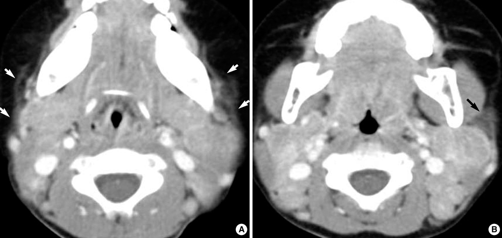
Fig. 2 A two-year-old girl with fever and bilateral cervical lymphadenopathy. Axial CT scans show multiple, medium-dimension lymph nodes with uniform enhancement on both sides of the neck at levels II and V. Note the increased attenuation in the adjacent superficial space (arrows).
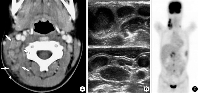
Fig. 3 A 10-yr-old girl with intermittent fever and right cervical lymphadenopathy. (A) CT scan shows multiple, small- and medium-sized lymph nodes on the right side of the neck (arrows) (levels IIB and V). Note the perinodal fat obliteration in the adjacent superficial space (arrowhead). (B) US obtained at the same time show somewhat matted, hypoechoic, nodal masses with irregular perinodal borders, suggestive of perinodal infiltrates. (C) MIP FDG-PET scan image shows multiple areas of FDG uptake in the right cervical, supraclavicular, right para-aortic and peripancreatic regions.
Reference
-
1. Lee KY, Yeon YH, Lee BC. Kikuchi-Fujimoto disease with prolonged fever in children. Pediatrics. 2004. 114:e752–e756.
Article2. Lin HC, Su CY, Huang SC. Kikuchi's disease in Asian children. Pediatrics. 2005. 115:e92–e96.3. Som PM, Curtin HD, Mancuso AA. Imaging-based nodal classification for evaluation of neck metastatic adenopathy. AJR Am J Roentgenol. 2000. 174:837–844.
Article4. Tsang WY, Chan JK, Ng CS. Kikuchi's lymphadenitis. A morphologic analysis of 75 cases with special reference to unusual features. Am J Surg Pathol. 1994. 18:219–231.5. Kwon SY, Kim TK, Kim YS, Lee KY, Lee NJ, Seol HY. CT findings in Kikuchi disease: analysis of 96 cases. AJNR Am J Neuroradiol. 2004. 25:1099–1102.6. Turner RR, Martin J, Dorfman RF. Necrotizing lymphadenitis. A study of 30 cases. Am J Surg Pathol. 1983. 7:115–123.7. Miller WT Jr, Perez-Jaffe LA. Cross-sectional imaging of Kikuchi disease. J Comput Assist Tomogr. 1999. 23:548–551.
Article8. Pyeun YS, Lee SW, Rho MH. Ultrasonographic evaluation of Kikuchi disease in the neck. J Korean Soc Med Ultrasound. 2001. 20:221–226.9. Kaicker S, Gerard PS, Kalburgi S, Geller MD, Hailoo D. PET-CT scan in a patient with Kikuchi disease. Pediatr Radiol. 2008. 38:596–597.
Article
- Full Text Links
-
- Actions
-
Cited
- CITED
-
- Close
- Share
-
- Similar articles
-
- A case of systemic lupus erythematosus associated with Kikuchi's disease
- A Case of Kikuchi's Disease (Histiocytic Necrotizing Lymphadenopathy) Showing Typical Histopathologic Findings in Cutaneous Lesion
- Histiocytic Necrotizing Lymphadenitis(Kikuchi's Disease) A clinicopathologic study of 1 cases
- Clinical characteristics of Kikuchi disease in children
- Kikuchi-Fujimoto Disease, A Possible Complication of Rituximab Treatment
