The Time between Paraquat Ingestion and a Negative Dithionite Urine Test in an Independent Risk Factor for Death and Organ Failure in Acute Paraquat Intoxication
- Affiliations
-
- 1Department of Internal Medicine, Soonchunhyang University College of Medicine, Cheonan, Korea. syhong@sch.ac.kr
- 2Department of Immunology, Soonchunhyang University College of Medicine, Cheonan, Korea.
- KMID: 1782130
- DOI: http://doi.org/10.3346/jkms.2012.27.9.993
Abstract
- To identify a prognostic marker that is less sensitive to variations in the elapsed time since paraquat ingestion, we assessed the time between paraquat ingestion and a negative dithionite urine test as a prognostic parameter in patients with acute paraquat intoxication. Forty-one patients with acute paraquat intoxication were enrolled in this study and analyzed to verify significant determinants of mortality and organ dysfunction. The amount of paraquat ingested, paraquat plasma levels, and the time to a negative urine dithionite test were significant independent risk factors predicting mortality. The amount of paraquat ingestion, and the time to a negative urine dithionite test were independent risk factors predicting organ dysfunction. With a cut-off value of 34.5 hr for the time to negative conversion of the urine dithionite test, the sensitivity and specificity for mortality were 71.4% and 75.0%, respectively. The incidence of acute kidney injury and respiratory failure above 34.5 hr were 100% and 85.0%, respectively. In conclusion, the time to a negative urine dithionite test is the reliable marker for predicting mortality and/or essential organ failure in patients with acute paraquat intoxication, who survive 72 hr.
MeSH Terms
Figure
-

Fig. 1 Plasma paraquat levels of two patients who ingested paraquat. The case-1, 48-yr-old man, arrived at hospital 5 hr after 200 mL of paraquat ingestion and the initial plasma paraquat level was 29.8 µg/mL. He discharged moribundly to other hospital in the second hospital day. The case-2, 58-yr-old man, arrived 6 hr after 50 mL of paraquat ingestion and the initial plasma paraquat level was 4.2 µg/mL. He died 72 hr after paraquat intoxication because of cardiac arrest and acute respiratory failure. The plasma paraquat levels of both patients demonstrated transient increases within 7 hr of paraquat ingestion followed by an abrupt drop. The plasma concentrations demonstrate substantial variations in concentration even with slight variations in the time interval since ingestion.
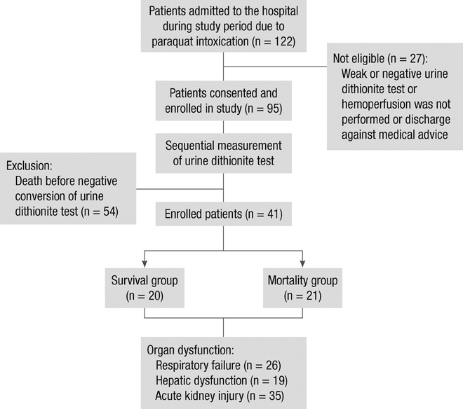
Fig. 2 Patient enrollment diagram in this study.
Reference
-
1. Gawarammana IB, Buckley NA. Medical management of paraquat ingestion. Br J Clin Pharmacol. 2011. 72:745–757.2. Lin JL, Lin-Tan DT, Chen KH, Huang WH, Hsu CW, Hsu HH, Yen TH. Improved survival in severe paraquat poisoning with repeated pulse therapy of cyclophosphamide and steroids. Intensive Care Med. 2011. 37:1006–1013.3. Moon JM, Chun BJ. The efficacy of high doses of vitamin C in patients with paraquat poisoning. Hum Exp Toxicol. 2011. 30:844–850.4. Kim JH, Gil HW, Yang JO, Lee EY, Hong SY. Effect of glutathione administration on serum levels of reactive oxygen metabolites in patients with paraquat intoxication: a pilot study. Korean J Intern Med. 2010. 25:282–287.5. Gil HW, Kim SJ, Yang JO, Lee EY, Hong SY. Clinical outcome of hemoperfusion in poisoned patients. Blood Purif. 2010. 30:84–88.6. Suzuki K, Takasu N, Arita S, Maenosono A, Ishimatsu S, Nishina M, Tanaka S, Kohama A. A new method for predicting the outcome and survival period in paraquat poisoning. Hum Toxicol. 1989. 8:33–38.7. Ragoucy-Sengler C, Pileire B. A biological index to predict patient outcome in paraquat poisoning. Hum Exp Toxicol. 1996. 15:265–268.8. Yamaguchi H, Sato S, Watanabe S, Naito H. Pre-embarkment prognostication for acute paraquat poisoning. Hum Exp Toxicol. 1990. 9:381–384.9. Ikebuchi J, Proudfoot AT, Matsubara K, Hampson EC, Tomita M, Suzuki K, Fuke C, Ijiri I, Tsunerari T, Yuasa I. Toxicological index of paraquat: a new strategy for assessment of severity of paraquat poisoning in 128 patients. Forensic Sci Int. 1993. 59:85–87.10. Proudfoot AT, Stewart MS, Levitt T, Widdop B. Significance of plasma-paraquat concentrations. Lancet. 1979. 2:330–332.11. Gil HW, Kang MS, Yang JO, Lee EY, Hong SY. Association between plasma paraquat level and outcome of paraquat poisoning in 375 paraquat poisoning patients. Clin Toxicol (Phila). 2008. 46:515–518.12. Lee KH, Gil HW, Kim YT, Yang JO, Lee EY, Hong SY. Marked recovery from paraquat-induced lung injury during long-term follow-up. Korean J Intern Med. 2009. 24:95–100.13. Kim YT, Jou SS, Lee HS, Gil HW, Yang JO, Lee EY, Hong SY. The area of ground glass opacities of the lungs as a predictive factor in acute paraquat intoxication. J Korean Med Sci. 2009. 24:636–640.14. Kim SJ, Gil HW, Yang JO, Lee EY, Hong SY. The clinical features of acute kidney injury in patients with acute paraquat intoxication. Nephrol Dial Transplant. 2009. 24:1226–1232.15. Gil HW, Yang JO, Lee EY, Hong SY. Clinical implication of urinary neutrophil gelatinase-associated lipocalin and kidney injury molecule-1 in patients with acute paraquat intoxication. Clin Toxicol (Phila). 2009. 47:870–875.16. Shivhare P, Gupta VK. Spectrophotometric method for the determination of paraquat in water, grain and plant materials. Analyst. 1991. 116:391–393.17. de Almeida RM, Yonamine M. Enzymatic-spectrophotometric determination of paraquat in urine samples: a method based on its toxic mechanism. Toxicol Mech Methods. 2010. 20:424–427.18. Braithwaite RA. Emergency analysis of paraquat in biological fluids. Hum Toxicol. 1987. 6:83–86.19. Kawase S, Kanno S, Skai S. Determination of the herbicides paraquat and diquat in blood and urine by gas chromatography. J Chromatogr. 1984. 283:231–240.20. Bowles MR, Eyles DW, Hampson EC, Pond SM. Quantitation of paraquat in biological samples by radioimmunoassay using a monoclonal antibody. Fundam Appl Toxicol. 1992. 19:375–379.21. Nagao M, Takatori T, Terazawa K, Wu B, Wakasugi C, Masui M, Ikeda H. Development and application of immunoassay for paraquat: radioimmunoassay. J Forensic Sci. 1989. 34:547–552.22. Mayhew SG. The redox potential of dithionite and SO-2 from equilibrium reactions with flavodoxins, methyl viologen and hydrogen plus hydrogenase. Eur J Biochem. 1978. 85:535–547.23. Houzé P, Baud FJ, Mouy R, Bismuth C, Bourdon R, Scherrmann JM. Toxicokinetics of paraquat in humans. Hum Exp Toxicol. 1990. 9:5–12.24. Kang MS, Gil HW, Yang JO, Lee EY, Hong SY. Comparison between kidney and hemoperfusion for paraquat elimination. J Korean Med Sci. 2009. 24:S156–S160.
- Full Text Links
-
- Actions
-
Cited
- CITED
-
- Close
- Share
-
- Similar articles
-
- Comparative Study Evaluating the Accuracy of the Urine Sodium Dithionite Test to Predict Plasma Paraquat Concentration in Poisoning Cases
- Usefulness of Hemoperfusion in Paraquat Poisoning
- Hart's survival curve are Very Useful for Predicting Prognosis in Paraquat Poisoning
- Plasma Paraquat Concentration and the Severity Index of Paraquat Poisoning (SIPP) at Presentation in Paraquat Intoxication
- Clinical Observation of Paraquat Poisoning
