Diffusion-Weighted Imaging with Sensitivity Encoding (SENSE) for Detecting Cranial Bone Marrow Metastases: Comparison with T1-Weighted Images
- Affiliations
-
- 1Department of Radiology, Kangbuk Samsung Hospital, Sungkyunkwan University School of Medicine, Seoul, Korea. ec.chung@samsung.com
- 2Department of Radiology, Konkuk University Hospital, Konkuk University School of Medicine, Seoul , Korea.
- KMID: 1779432
- DOI: http://doi.org/10.3348/kjr.2007.8.3.185
Abstract
OBJECTIVE
This study was designed to determine whether diffusion-weighted imaging (DWI) with sensitivity encoding (SENSE) could detect bone marrow involvement in patients with cranial bone marrow (CBM) metastases. DWI results obtained were compared with T1-weighted imaging (T1WI) findings. MATERIALS AND METHODS: DWI with sensitivity encoding (SENSE; b value = 1,000) was performed consecutively in 13 patients with CBM metastases diagnosed pathologically and radiologically. CBM lesions were dichotomized according to the involved site, i.e., skull base or calvarium. Two radiologists qualitatively evaluated the relative conspicuousness of CBM lesions and image qualities in B0 and in isotropic DWI and in T1WI. According to region of interest analysis of normal and pathologic marrow for these three sequences, absolute signal difference percentages (SD%) were calculated to quantitatively analyze lesion contrast. RESULTS: All 20 lesions in 13 patients with CBM metastases revealed abnormal DWI signals in areas corresponding to T1WI abnormalities. Both skull base and calvarial lesions provided better lesion conspicuousness than T1WI and B0 images. Although the image quality of DWI was less satisfactory than that of T1WI, relatively good image qualities were obtained. Quantitatively, B0 images (SD%, 82.1+/-7.9%) showed better lesion contrast than isotropic DWI (SD%, 71.4+/-13.7%) and T1WI (SD%, 65.7+/-9.3%) images. CONCLUSION: For scan times of less than 30 seconds, DWI with SENSE was able to detect bone marrow involvement, and was superior to T1WI in terms of lesion conspicuity. DWI with SENSE may be helpful for the detection of cranial bone/bone marrow metastases when used in conjunction with conventional MR sequences.
Keyword
MeSH Terms
Figure
-
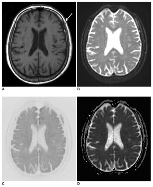
Fig. 1 A 71-year-old woman with lung cancer (case no. 4). T1-weighted image (A), B0 image (B), isotropic diffusion-weighted image (C), and an apparent diffusion coefficient map (D) were obtained. Abnormalities in signal intensity due to bone marrow metastases (arrow) are evident on all images. However, the relative lesion conspicuousness is better on the B0 image and the isotropic diffusion-weighted image than on the T1-weighted image.
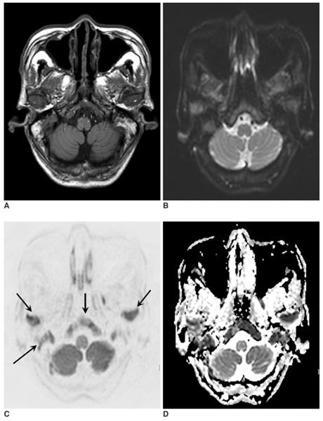
Fig. 2 A 69-year-old man with lung cancer (case no. 3). T1-weighted image (A), B0 image (B), isotropic diffusion-weighted image (C), and an apparent diffusion-coefficient map (D) were obtained. Lesions involving the skull base and mandibular condyles (arrows) are visible on all images. However, the isotropic diffusion-weighted image shows a superior lesion contrast.
Reference
-
1. Yildirim T, Agildere AM, Oguzkurt L, Barutcu O, Kizilkilic O, Kocak R, et al. MRI evaluation of cranial bone marrow signal intensity and thickness in chronic anemia. Eur J Radiol. 2005. 53:125–130.2. Loevner LA, Tobey JD, Yousem DM, Sonners AI, Hsu WC. MR imaging characteristics of cranial bone marrow in adult patients with underlying systemic disorders compared with healthy control subjects. AJNR Am J Neuroradiol. 2002. 23:248–254.3. Vogler JB 3rd, Murphy WA. Bone marrow imaging. Radiology. 1988. 168:679–693.4. Daffner RH, Lupetin AR, Dash N, Deeb ZL, Sefczek RJ, Schapiro RL. MRI in the detection of malignant infiltration of bone marrow. AJR Am J Roentgenol. 1986. 146:353–358.5. Nyman R, Rehn S, Glimelius B, Hagberg H, Hemmingsson A, Jung B, et al. Magnetic resonance imaging in diffuse malignant bone marrow disease. Acta Radiol. 1987. 28:199–205.6. Ricci C, Cova M, Kang YS, Yang A, Rahmouni A, Scott WW Jr, et al. Normal age-related patterns of cellular and fatty bone marrow distribution in the axial skeleton: MR imaging study. Radiology. 1990. 177:83–88.7. Herneth AM, Friedrich K, Weidekamm C, Schibany N, Krestan C, Czerny C, et al. Diffusion weighted imaging of bone marrow pathologies. Eur J Radiol. 2005. 55:74–83.8. Raya JG, Dietrich O, Reiser MF, Baur-Melnyk A. Techniques for diffusion-weighted imaging of bone marrow. Eur J Radiol. 2005. 55:64–73.9. Baur A, Stabler A, Bruning R, Bartl R, Krodel A, Reiser M, et al. Diffusion-weighted MR imaging of bone marrow: differentiation of benign versus pathologic compression fractures. Radiology. 1998. 207:349–356.10. Castillo M, Arbelaez A, Smith JK, Fisher LL. Diffusion-weighted MR imaging offers no advantage over routine noncontrast MR imaging in the detection of vertebral metastases. AJNR Am J Neuroradiol. 2000. 21:948–953.11. Herneth AM, Philipp MO, Naude J, Funovics M, Beichel RR, Bammer R, et al. Vertebral metastases: assessment with apparent diffusion coefficient. Radiology. 2002. 225:889–894.12. Maeda M, Sakuma H, Maier SE, Takeda K. Quantitative assessment of diffusion abnormalities in benign and malignant vertebral compression fractures by line scan diffusion-weighted imaging. AJR Am J Roentgenol. 2003. 181:1203–1209.13. Zhou XJ, Leeds NE, McKinnon GC, Kumar AJ. Characterization of benign and metastatic vertebral compression fractures with quantitative diffusion MR imaging. AJNR Am J Neuroradiol. 2002. 23:165–170.14. Byun WM, Shin SO, Chang Y, Lee SJ, Finsterbusch J, Frahm J. Diffusion-weighted MR imaging of metastatic disease of the spine: assessment of response to therapy. AJNR Am J Neuroradiol. 2002. 23:906–912.15. Meyer JR, Gutierrez A, Mock B, Hebron D, Prager JM, Gorey MT, et al. High-b-value diffusion-weighted MR imaging of suspected brain infarction. AJNR Am J Neuroradiol. 2000. 21:1821–1829.16. Mirowitz SA, Apicella P, Reinus WR, Hammerman AM. MR imaging of bone marrow lesions: relative conspicuousness on T1-weighted, fat-suppressed T2-weighted, and STIR images. AJR Am J Roentgenol. 1994. 162:215–221.17. Stadnik TW, Chaskis C, Michotte A, Shabana WM, van Rompaey K, Luypaert R, et al. Diffusion-weighted MR imaging of intracerebral masses: comparison with conventional MR imaging and histologic findings. AJNR Am J Neuroradiol. 2001. 22:969–976.18. Takahara T, Imai Y, Tamashita T, Yasuda S, Nasu S, Van Cauteren M. Diffusion-weighted whole body imaging with background body signal suppression (DWIBS): technical improvement using free breathing, STIR and high resolution 3D display. Radiat Med. 2004. 22:275–282.19. Bammer R, Keeling SL, Augustin M, Pruessmann KP, Wolf R, Stollberger R, et al. Improved diffusion-weighted single-shot echo-planar imaging (EPI) in stroke using sensitivity encoding (SENSE). Magn Reson Med. 2001. 46:548–554.20. Chan JH, Peh WC, Tsui EY, Chau LF, Cheung KK, Chan KB, et al. Acute vertebral body compression fractures: discrimination between benign and malignant causes using apparent diffusion coefficients. Br J Radiol. 2002. 75:207–214.21. Haubold-Reuter BG, Duewell S, Schilcher BR, Marincek B, von Schulthess GK. The value of bone scintigraphy, bone marrow scintigraphy and fast spin-echo magnetic resonance imaging in staging of patients with malignant solid tumours: a prospective study. Eur J Nucl Med. 1993. 20:1063–1069.22. Daldrup-Link HE, Franzius C, Link TM, Laukamp D, Sciuk J, Jurgens H, et al. Whole-body MR imaging for detection of bone metastases in children and young adults: comparison with skeletal scintigraphy and FDG PET. AJR Am J Roentgenol. 2001. 177:229–236.23. Hamaoka T, Madewell JE, Podoloff DA, Hortobagyi GN, Ueno NT. Bone imaging in metastatic breast cancer. J Clin Oncol. 2004. 22:2942–2953.
- Full Text Links
-
- Actions
-
Cited
- CITED
-
- Close
- Share
-
- Similar articles
-
- The Usefulness of Diffusion-weighted MR Imaging for Differentiation between Degenerative Spines and Infectious Spondylitis
- The Role of Diffusion-Weighted Imaging and the Apparent Diffusion Coefficient (ADC) Values for Breast Tumors
- Evaluation of Treatment Response Using Diffusion-Weighted MRI in Metastatic Spines
- The Value of PROPELLER Diffusion-Weighted Image in the Detection of Cholesteatoma
- Abbreviated Breast Magnetic Resonance Imaging: Background, Evidence From Studies, and Future Considerations
