The Role of Diffusion-Weighted Imaging and the Apparent Diffusion Coefficient (ADC) Values for Breast Tumors
- Affiliations
-
- 1Department of Radiology, St. Vincent's Hospital, The Catholic University of Korea, Suwon, Korea. escha@catholic.ac.kr
- KMID: 1734287
- DOI: http://doi.org/10.3348/kjr.2007.8.5.390
Abstract
OBJECTIVE
We wanted to evaluate the role of diffusion-weighted imaging (DWI) and the apparent diffusion coefficient (ADC) for detecting breast tumors, as compared with the T1- and T2-weighted images. MATERIALS AND METHODS: Forty-one female patients underwent breast MRI, and this included the T1-, T2-, DWI and dynamic contrast-enhanced images. Sixty-five enhancing lesions were detected on the dynamic contrast-enhanced images and we used this as a reference image for detecting tumor. Fifty-six breast lesions were detected on DWI and the histological diagnoses were as follows: 43 invasive ductal carcinomas, one mucinous carcinoma, one mixed infiltrative and mucinous carcinoma, seven ductal carcinomas in situ (DCIS), and four benign tumors. First, we compared the detectability of breast lesions on DWI with that of the T1- and T2-weighted images. We then compared the ADCs of the malignant and benign breast lesions to the ADCs of the normal fibroglandular tissue. RESULTS: Fifty-six lesions were detected via DWI (detectability of 86.2%). The detectabilities of breast lesions on the T1- and T2-weighted imaging were 61.5% (40/65) and 75.4% (49/65), respectively. The mean ADCs of the invasive ductal carcinoma (0.89 +/- 0.18x10(-3)mm2/second) and DCIS (1.17 +/- 0.18x10(-3)mm2/ second) are significantly lower than those of the benign lesions (1.41 +/- 0.56x10(-3)mm2/second) and the normal fibroglandular tissue (1.51 +/- 0.29x10(-3)mm2/ second). CONCLUSION: DWI has a high sensitivity for detecting breast tumors, and especially for detecting malignant breast tumors. DWI was an effective imaging technique for detecting breast lesions, as compared to using the T1- and T2-weighted images.
MeSH Terms
-
Adenocarcinoma, Mucinous/*diagnosis
Adult
Aged
Breast/pathology
Breast Neoplasms/*diagnosis
Carcinoma, Ductal, Breast/*diagnosis
Carcinoma, Intraductal, Noninfiltrating/*diagnosis
Contrast Media/administration & dosage
Diffusion Magnetic Resonance Imaging/*methods
Female
Gadolinium DTPA/diagnostic use
Humans
Image Enhancement/methods
Imaging, Three-Dimensional/methods
Middle Aged
Observer Variation
Sensitivity and Specificity
Figure
-
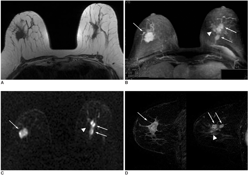
Fig. 1 A 69-year-old woman with bilateral invasive ductal carcinoma. A.The axial T1-weighted image shows multiple lobular homogeneous iso-signal intensity masses in both breasts. B.The maximum-intensity projection of the subtraction image shows multiple enhancing masses in both breasts (thin white arrows and arrowhead). C.The axial diffusion weighted image shows multiple high-signal intensity masses in both breasts (thin white arrows), but tiny multicentric lesions are not detected in the left breast (white arrowhead). The apparent difusion coefficient value of the right breast tumor was 0.94 × 10⁻³mm2/second and that of the left breast tumor was 0.84 × 10⁻³mm2/second. D.The sagittal dynamic-enhanced T1-weighted gradient-echo substraction image of the first postcontrast acquisition shows multiple, heterogeneous, rim-enhancing masses in both breasts (thin white arrows and white arrowhead). The left side of this image represents the right breast and the right one represents the left breast. A tiny multicentric lesion is extremely well visualized in the left breast (white arrowhead).

Fig. 2 A 53-year-old woman with ductal carcinoma in situ. A.The axial T1-weighted image shows segmentally distributed, asymmetric, iso-signal intensity, non-mass lesion in the left breast. B.The maximal intensity projection of a subtraction image shows heterogeneous clumpy enhancement in the left breast. C.The diffusion-weighted image shows the main lesion to be a high-signal intensity lesion. D.The axial plane apparent diffusion coefficient map shows a mixed green and yellow area and the apparent diffusion coefficient value of this breast tumor was 1.43 × 10⁻³mm2/second.

Fig. 3 A 41-year-old woman with fibroadenoma that was confirmed by core-needle biopsy. A.The axial T1-weighted image shows a circumscribed oval iso-signal intensity mass in the right breast. B.The maximal intensity projection of the subtraction image shows homogeneous enhancement in the right breast. A nonspecific small enhancing nodule is also seen in the left breast. This left nodule showed typical benign features on the breast USG (not shown), so we did not perform core-needle biopsy. C.The diffusion-weighted image shows the mass to be a high-signal intensity lesion in the right breast. D.The axial plane apparent diffusion coefficient map shows the mixed yellow and red area, and the apparent diffusion coefficient value of this tumor was 1.63 × 10⁻³mm2/second.
Cited by 1 articles
-
Correlation of Prognostic Factors of Invasive Lobular Carcinoma with ADC Value of DWI and SUVMax of FDG-PET
Bo Bae Choi, Sung Hun Kim, Chang Suk Park, Na Young Jung
Chonnam Med J. 2017;53(2):133-139. doi: 10.4068/cmj.2017.53.2.133.
Reference
-
1. Bammer R. Basic principles of diffusion-weighted imaging. Eur J Radiol. 2003. 45:169–184.2. Guo Y, Cai YQ, Cai ZL, Gao YG, An NY, Ma L, et al. Differentiation of clinically benign and malignant breast lesions using diffusion-weighted imaging. J Magn Reson Imaging. 2002. 16:172–178.3. Sinha S, Lucas-Quesada FA, Sinha U, DeBruhl N, Bassett LW. In vivo diffusion-weighted MRI of the breast: potential for lesion characterization. J Magn Reson Imaging. 2002. 15:693–704.4. Kinoshita T, Yashiro N, Ihara N, Funatu H, Fakuma E, Narita M. Diffusion-weighted half-Fourier single-shot turbo spin echo imaging in breast tumor: differentiation of invasive ductal carcinoma from fibroadenoma. J Comput Assist Tomogr. 2002. 26:1042–1046.5. Woodhams R, Matsunaga K, Iwabuchi K, Kan S, Hata H, Kuranami M, et al. Diffusion-weighted imaging of malignant breast tumors: the usefulness of apparent diffusion coefficient (ADC) value and ADC map for the detection of malignant breast tumors and evaluation of cancer extension. J Comput Assist Tomogr. 2005. 29:644–649.6. Kim T, Murakami T, Takahashi S, Tsuda K, Nakamura H. Diffusion-weighted single-shot echoplanar MR Imaging for liver disease. AJR Am J Roentgenol. 1999. 173:393–398.7. Ichikawa T, Haradome H, Hachiya J, Nitatori T, Araki T. Diffusion-weighted MR imaging with a single-shot echoplanar sequence: detection and characterization of focal hepatic lesions. AJR Am J Roentgenol. 1998. 170:397–402.8. Yamashita Y, Namimoto T, Mitsuzaki K, Urata J, Tsuchigame T, Takahashi M, et al. Mucin-producing tumor of the pancreas: diagnostic value of diffusion-weighted echo-planar MRI imaging. Radiology. 1998. 208:605–609.9. Moteki T, Ishizaka H. Diffusion-weighted EPI of cystic ovarian lesions: evaluation of cystic contents using apparent diffusion coefficients. J Magn Reson Imaging. 2000. 12:1014–1019.10. Hosseinzadeh K, Schwarz SD. Endorectal diffusion-weighted imaging in prostate cancer to differentiate malignant and benign peripheral zone tissue. J Magn Reson Imaging. 2004. 20:654–661.11. Bluemke DA, Gatsonis CA, Chen MH, DeAngelis GA, DeBruhl N, Harms S, et al. Magnetic resonance imaging of the breast prior to biopsy. JAMA. 2004. 292:2735–2742.12. Orel SG, Schnall MD. MR imaging of the breast for the detection, diagnosis, and staging of breast cancer. Radiology. 2001. 220:13–30.13. DeBruhl ND, Michael D, Bassett LW. Bassett LW, Jackson VP, Fu KL, Fu YS, editors. Magnetic resonance imaging of breast tumors. Diagnosis of diseases of the breast. 2005. 2nd ed. Philadelphia: Elsevier Saunders;225–250.14. Ikeda DM, Baker DR, Daniel BL. Magnetic resonance imaging of breast cancer : clinical indications and breast MRI reporting system. J Magn Reson Imaging. 2000. 12:975–983.15. Siegmann KC, Muller-Schimpfle M, Schick F, Remy CT, Fersis N, Ruck P, et al. MR imaging-detected breast lesion: histopathologic correlation of lesion characteristics and signal intensity data. AJR Am J Roentgenol. 2002. 178:1403–1409.16. Jacobs MA, Barker PB, Bluemke DA, Maranto C, Arnold C, Herskovits EH, et al. Benign and malignant breast lesions: diagnosis with multiparametric MR imaging. Radiology. 2003. 229:225–232.17. Warach S, Boska M, Welch KM. Pitfalls and potential of clinical diffusion-weighted MR imaging in acute stroke. Stroke. 1997. 28:481–482.18. Sugahara T, Korogi Y, Kochi M, Ikushima I, Shigematu Y, Hirai T, et al. Usefulness of diffusion-weighted MRI with echo-planar technique in the evaluation of cellularity in gliomas. J Magn Reson Imaging. 1999. 9:53–60.
- Full Text Links
-
- Actions
-
Cited
- CITED
-
- Close
- Share
-
- Similar articles
-
- Reversal of a Large Ischemic Lesion with Low Apparent Diffusion Coefficient Value by Rapid Spontaneous Recanalization
- Clinical applications and characteristics of apparent diffusion coefficient maps for the brain of two dogs
- Usefulness of Apparent Diffusion Coefficient in Ovarian Cystic Tumors Using Diffusion-Weighted Magnetic Resonance Imaging
- Diffusion-weighted Imaging and Apparent Diffusion Coefficient Maps for the Evaluation of Pyogenic Ventriculitis
- Usefulness of Diffusion-Weighted MR Imaging for Breast Lesions: Comparing the Apparent Diffusion Coefficient (ADC) Values and the Pathologic Results
