Yonsei Med J.
2012 May;53(3):654-657. 10.3349/ymj.2012.53.3.654.
Phagocytosis and Endocytosis of Silver Nanoparticles Induce Interleukin-8 Production in Human Macrophages
- Affiliations
-
- 1Department of Microbiology, Brain Korea 21 Project for Medical Science, Institute for Immunology and Immunological Diseases, Yonsei University College of Medicine, Seoul, Korea. inhong@yuhs.ac
- KMID: 1777004
- DOI: http://doi.org/10.3349/ymj.2012.53.3.654
Abstract
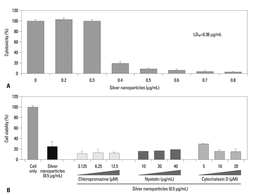
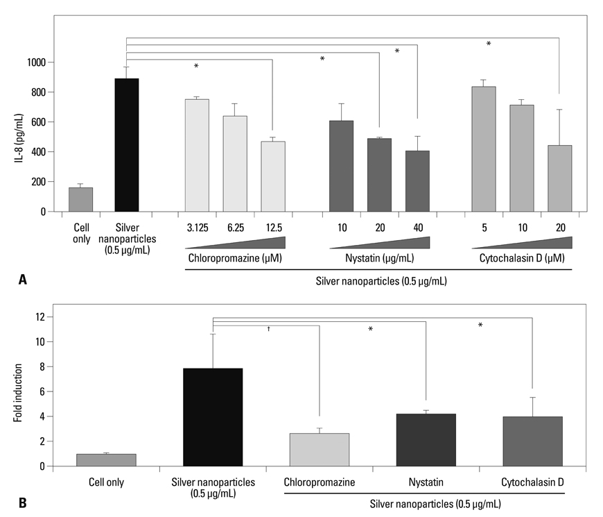
- Phagocytosis or endocytosis by macrophages is critical to the uptake of fine particles, including nanoparticles, in order to initiate toxic effects in cells. Here, our data enhance the understanding of the process of internalization of silver nanoparticles by macrophages. When macrophages were pre-treated with inhibitors to phagocytosis, caveolin-mediated endocytosis, or clathrin-mediated endocytosis, prior to exposure to silver nanoparticles, Interleukin-8 (IL-8) production was inhibited. Although cell death was not reduced, the inflammatory response by macrophages was compromised by phagocytosis and endocytosis inhibitors.
Keyword
MeSH Terms
Figure
Cited by 2 articles
-
The Effects of Silica Nanoparticles in Macrophage Cells
Seungjae Kim, Jiyoung Jang, Hyojin Kim, Hoon Choi, Kangtaek Lee, In-Hong Choi
Immune Netw. 2012;12(6):296-300. doi: 10.4110/in.2012.12.6.296.The Effects of Silica Nanoparticles in Macrophage Cells
Seungjae Kim, Jiyoung Jang, Hyojin Kim, Hoon Choi, Kangtaek Lee, In-Hong Choi
Immune Netw. 2012;12(6):296-300. doi: 10.4110/in.2012.12.6.296.
Reference
-
1. Yokel RA, Macphail RC. Engineered nanomaterials: exposures, hazards, and risk prevention. J Occup Med Toxicol. 2011. 6:7.
Article2. Hackenberg S, Scherzed A, Kessler M, Hummel S, Technau A, Froelich K, et al. Silver nanoparticles: evaluation of DNA damage, toxicity and functional impairment in human mesenchymal stem cells. Toxicol Lett. 2011. 201:27–33.
Article3. Jang J, Lim DH, Choi IH. The impact of nanomaterials in immune system. Immune Netw. 2010. 10:85–91.
Article4. Tantra R, Knight A. Cellular uptake and intracellular fate of engineered nanoparticles: a review on the application of imaging techniques. Nanotoxicology. 2011. 5:381–392.
Article5. Zhao F, Zhao Y, Liu Y, Chang X, Chen C, Zhao Y. Cellular uptake, intracellular trafficking, and cytotoxicity of nanomaterials. Small. 2011. 7:1322–1337.
Article6. Kerr MC, Teasdale RD. Defining macropinocytosis. Traffic. 2009. 10:364–371.
Article7. Kumari S, Mg S, Mayor S. Endocytosis unplugged: multiple ways to enter the cell. Cell Res. 2010. 20:256–275.
Article8. Hernaez B, Alonso C. Dynamin- and clathrin-dependent endocytosis in African swine fever virus entry. J Virol. 2010. 84:2100–2109.
Article9. Santibanez JF, Blanco FJ, Garrido-Martin EM, Sanz-Rodriguez F, del Pozo MA, Bernabeu C. Caveolin-1 interacts and cooperates with the transforming growth factor-beta type I receptor ALK1 in endothelial caveolae. Cardiovasc Res. 2008. 77:791–799.
Article10. Greulich C, Diendorf J, Simon T, Eggeler G, Epple M, Köller M. Uptake and intracellular distribution of silver nanoparticles in human mesenchymal stem cells. Acta Biomater. 2011. 7:347–354.
Article11. Wang T, Bai J, Jiang X, Nienhaus GU. Cellular uptake of nanoparticles by membrane penetration: a study combining confocal microscopy with FTIR spectroelectrochemistry. ACS Nano. 2012. 6:1251–1259.
Article12. Dubavik A, Sezgin E, Lesnyak V, Gaponik N, Schwille P, Eychmüller A. Penetration of amphiphilic quantum dots through model and cellular plasma membranes. ACS Nano. 2012. 02. 13. [Epub ahead of print].
Article13. Lin J, Zhang H, Chen Z, Zheng Y. Penetration of lipid membranes by gold nanoparticles: insights into cellular uptake, cytotoxicity, and their relationship. ACS Nano. 2010. 4:5421–5429.
Article14. Oh E, Delehanty JB, Sapsford KE, Susumu K, Goswami R, Blanco-Canosa JB, et al. Cellular uptake and fate of PEGylated gold nanoparticles is dependent on both cell-penetration peptides and particle size. ACS Nano. 2011. 5:6434–6448.
Article15. Park J, Lim DH, Lim HJ, Kwon T, Choi JS, Jeong S, et al. Size dependent macrophage responses and toxicological effects of Ag nanoparticles. Chem Commun (Camb). 2011. 47:4382–4384.
Article16. Zhang A, Guan Y, Xu LX. Theoretical study on temperature dependence of cellular uptake of QDs nanoparticles. J Biomech Eng. 2011. 133:124502.
Article17. Zhang LW, Bäumer W, Monteiro-Riviere NA. Cellular uptake mechanisms and toxicity of quantum dots in dendritic cells. Nanomedicine (Lond). 2011. 6:777–791.
Article
- Full Text Links
- Actions
-
Cited
- CITED
-
- Close
- Share
- Similar articles
-
- The Effects of Silica Nanoparticles in Macrophage Cells
- Silver Nanoparticles as a Smart Antimicrobial Agent
- Immunostimulatory Effects of Silica Nanoparticles in Human Monocytes
- Application of Silver Nanoparticles for the Control of Colletotrichum Species In Vitro and Pepper Anthracnose Disease in Field
- Effects of Interleukin 4 on the Production of Interleukin 6 in Human Keratinocytes


