Korean Circ J.
2008 Sep;38(9):468-474. 10.4070/kcj.2008.38.9.468.
Assessment of Intermediate Coronary Stenosis in Koreans Using the Fractional Flow Reserve
- Affiliations
-
- 1Division of Cardiology, Department of Internal Medicine, Seoul National University College of Medicine, Cardiovascular Center, Seoul National University Hospital, Seoul, Korea. bkkoo@snu.ac.kr
- 2Cardiovascular Center, Seoul National University Bundang Hospital, Seongnam, Korea.
- 3Department of Internal Medicine, Seoul National University College of Medicine, Seoul Metropolitan Boramae Hospital, Seoul, Korea.
- KMID: 1776347
- DOI: http://doi.org/10.4070/kcj.2008.38.9.468
Abstract
- BACKGROUND AND OBJECTIVES
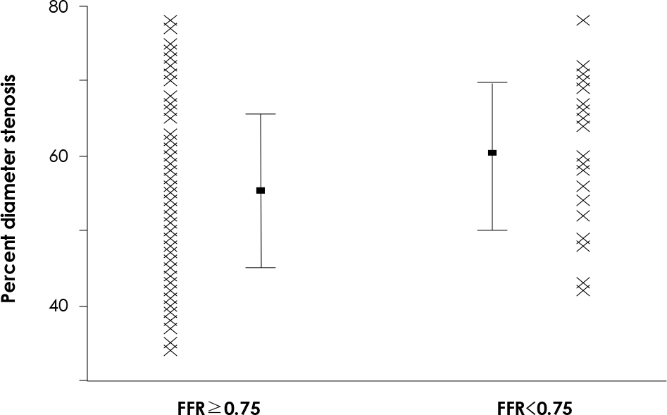
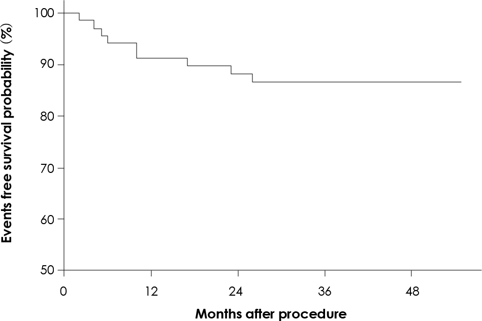
Previous studies that used physiologic parameters have shown that coronary angiography is not always accurate in the evaluation of intermediate lesions. We sought to evaluate the outcomes of Korean patients for whom revascularization was deferred according to their fractional flow reserve (FFR). SUBJECTS AND METHODS: FFR was measured in 107 intermediate lesions (visually estimated percent stenosis: 40-70%) in 102 consecutive patients (68% males, mean age: 62+/-9 years). The one-year cardiac adverse outcomes (cardiac death, myocardial infarction or target vessel revascularization) of all the patients and the long-term outcomes of the patients for whom revascularization was deferred according to the FFR were evaluated. RESULTS: The mean percent diameter stenosis was 57+/-11% and the FFR was 0.82+/-0.10. Only 25 lesions (23%) had a FFR <0.75. There was no significant difference in the 1-year cardiac event rates between the FFR > or =0.75 group and the FFR <0.75 group (10.4% vs. 13.0%, respectively, p=0.49). There was a tendency of a lower incidence of 1-year cardiac events in the medical treatment group than in the revascularizaton group (8.0% vs. 20.0%, respectively, p=0.10). In the 69 patients with FFR-guided deferral of revascularization, the long-term (mean follow-up duration: 41+/-11 months) cardiac event-free survival rate was 86%. CONCLUSION: The measurement of FFR seems to be a useful guide for decision making and it may reduce unnecessary intervention in Korean patients who suffer with intermediate stenosis.
MeSH Terms
Figure
Reference
-
References
1. Iskander S, Iskandrian AE. Risk assessment using single-photon emission computed tomographic technetium-99m sestamibi imaging. J Am Coll Cardiol. 1998; 32:57–62.
Article2. Gould KL, Lipscomb K, Hamilton GW. Physiologic basis for assessing critical coronary stenosis: instantaneous flow response and regional distribution during coronary hyperemia as measures of coronary flow reserve. Am J Cardiol. 1974; 33:87–94.3. De Rouen TA, Murray JA, Owen W. Variability in the analysis of coronary arteriograms. Circulation. 1977; 55:324–8.
Article4. Goldberg RK, Kleiman NS, Minor ST, Abukhalil J, Raizner AE. Comparison of quantitative coronary angiography to visual estimates of lesion severity pre and post PTCA. Am Heart J. 1990; 119:178–84.
Article5. Zir LM. Observer variability in coronary angiography. Int J Cardiol. 1983; 3:171–3.6. Topol EJ, Ellis SG, Cosgrove DM, et al. Analysis of coronary angioplasty practice in the United States with an insurance-claims data base. Circulation. 1993; 87:1489–97.
Article7. Lima RSL, Watson DD, Goode AR, et al. Incremental value of combined perfusion and function over perfusion alone by gated SPECT myocardial perfusion imaging for detection of severe three-vessel coronary artery disease. J Am Coll Cardiol. 2003; 42:64–70.
Article8. Mintz GS, Nissen SE, Anderson WD, et al. American College of Cardiology clinical expert consensus document on standards for acquisition, measurement and reporting of intravascular ultrasound studies (IVUS): a report of the American college of cardiology task force on clinical expert consensus documents. J Am Coll Cardiol. 2001; 37:1478–92.9. Pijls NH, van Son JA, Kirkeeide RL, De Bruyne B, Gould KL. Experimental basis of determining maximum coronary myocardial, and collateral blood flow by pressure measurements for assessing functional stenosis severity before and after percutaneous transluminal coronary angioplasty. Circulation. 1993; 87:1354–67.
Article10. Pijls NHJ, De Bruyne B, Peels K, et al. Measurement of fractional flow reserve to assess the functional severity of coronary-artery stenoses. N Engl J Med. 1996; 334:1703–8.
Article11. Bech GJ, De Bruyne B, Pijls NH, et al. Fractional flow reserve to determine the appropriateness of angioplasty in moderate stenosis: a randomized trial. Circulation. 2001; 103:2928–34.12. Koo BK, Kim CH, Na SH, et al. Intracoronary continuous adenosine infusion. Circ J. 2005; 69:908–12.13. Suh JW, Koo BK, Jo SH, et al. Optimal dosage and method of administration of adenosine for measuring the coronary flow reserve and the fractional flow reserve in Koreans. Korean Circ J. 2006; 36:300–7.
Article14. Fischer JJ, Samady H, McPherson JA, et al. Comparison between visual assessment and quantitative angiography versus fractional flow reserve for native coronary narrowings of moderate severity. Am J Cardiol. 2002; 90:210–5.
Article15. Koo BK, Kang HJ, Youn TJ, et al. Physiologic assessment of jailed side branch lesions using fractional flow reserve. J Am Coll Cardiol. 2005; 46:633–7.
Article16. Ziaee A, Parham WA, Herrmann SC, Stewart RE, Lim MJ, Kern MJ. Lack of relation between imaging and physiology in ostial coronary artery narrowings. Am J Cardiol. 2004; 93:1404–7.
Article17. Abizaid AS, Mintz GS, Mehran R, et al. Long-term follow-up after percutaneous transluminal coronary angioplasty was not performed based on intravascular ultrasound findings: Importance of lumen dimensions. Circulation. 1999; 100:256–61.18. Takagi A, Tsurumi Y, Ishii Y, Suzuki K, Kawana M, Kasanuki H. Clinical potential of intravascular ultrasound for physiological assessment of coronary stenosis: relationship between quantitative ultrasound tomography and pressure-derived fractional flow reserve. Circulation. 1999; 100:250–5.19. Briguori C, Anzuini A, Airoldi F, et al. Intravascular ultrasound criteria for the assessment of the functional significance of intermediate coronary artery stenoses and comparison with fractional flow reserve. Am J Cardiol. 2001; 87:136–41.
Article20. Kim DH, Kwan J, Seo JK, et al. Fractional flow reserve in coronary artery disease: comparison with intravascular ultrasound. Korean Circ J. 1999; 29:773–80.
Article21. Pijls NH, van Schaardenburgh P, Manoharan G, et al. Percutaneous coronary intervention of functionally nonsignificant ste-ntosis: 5-year follow-up of the DEFER study. J Am Coll Cardiol. 2007; 49:2105–11.22. Bae JH, Rihal CS, Lerman A. Tissue characterization of coronary plaques using intravascular ultrasound/virtual histology. Korean Circ J. 2006; 36:553–8.
Article23. Nissen SE, Tuzcu EM, Schoenhagen P, et al. Effect of intensive compared with moderate lipid-lowering therapy on progression of coronary atherosclerosis: a randomized controlled trial. JAMA. 2004; 291:1071–80.24. Nissen SE, Nicholls SJ, Sipahi I, et al. Effect of very high-intensity statin therapy on regression of coronary atherosclerosis. JAMA. 2006; 295:1556–65.
Article25. Moses JW, Stone GW, Nikolsky E, et al. Drug-eluting stents in the treatment of intermediate lesions: pooled analysis from four randomized trials. J Am Coll Cardiol. 2006; 47:2164–71.26. Lavi S, Rihal CS, Yang EH, et al. The effect of drug eluting stents on cardiovascular events in patients with intermediate lesions and borderline fractional flow reserve. Catheter Cardiovasc Interv. 2007; 70:523–31.
Article27. Stone GW, Moses JW, Ellis SG, et al. Safety and efficacy of sirolimus- and paclitaxel-eluting coronary stents. N Engl J Med. 2007; 356:998–1008.
Article28. Lagerqvist B, James SK, Stenestrand U, et al. Long-term outcomes with drug-eluting stents versus bare-metal stents in Sweden. N Engl J Med. 2007; 356:1009–19.
Article
- Full Text Links
- Actions
-
Cited
- CITED
-
- Close
- Share
- Similar articles
-
- Funtional significance of the intermediate lesion in a single coronary artery assessed by fractional flow reserve
- A Case of Successful Percutaneous Coronary Intervention by Fractional Flow Reserve and 13N-Ammonia Positron Emission Tomography
- Fractional Flow Reserve: The Past, Present and Future
- Cost-effectiveness of Fractional Flow Reserve Versus Intravascular Ultrasound to Guide Percutaneous Coronary Intervention: Results From the FLAVOUR Study
- Future Directions in Coronary CT Angiography: CT-Fractional Flow Reserve, Plaque Vulnerability, and Quantitative Plaque Assessment


