A Case of Successful Percutaneous Coronary Intervention by Fractional Flow Reserve and 13N-Ammonia Positron Emission Tomography
- Affiliations
-
- 1Department of Cardiovascular Medicine, Heart Center, Chonnam National University Hospital, Gwangju, Korea. myungho@chollian.net
- KMID: 2005734
- DOI: http://doi.org/10.12997/jla.2015.4.1.39
Abstract
- A 43-year-old male presented with effort angina. Ammonia positron emission tomography (PET) revealed reversible perfusion defect in left anterior descending artery (LAD) and left circumflex artery (LCX) territories with decreased coronary flow reserve. Coronary angiogram showed significant stenosis in proximal LAD and intermediate diffuse stenosis in LCX and right coronary artery (RCA). Fractional flow reserve (FFR) showed similar results with ammonia PET. After percutaneous coronary intervention for LAD and LCX, flow and pressure checked by PET and FFR showed improvement. Simultaneously use of ammonia PET and FFR could be useful for determining ischemia-inducible lesion especially in diffuse intermediate lesion with discrepancy between functional studies.
MeSH Terms
Figure
-

Fig. 1 13N-ammonia PET findings. (A) Reversible perfusion defect in anterior and lateral LV wall was revealed, (B) Coronary flow reserve was decreased in left anterior descending artery and left circumflex artery territory.

Fig. 2 Coronary angiography. (A) Left coronary angiogram reveals significant stenosis in proximal LAD, (B) Left coronary angiogram shows intermediate stenosis in proximal LCX, (C)(D) Right coronary angiogram shows intermediate stenosis in distal RCA.
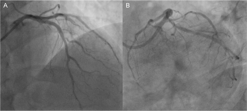
Fig. 3 Successful FFR and 13N-ammonia PET guided PCI with coronary stenting using drug-eluting stents (3.0×30 mm and 3.5×34 mm Resolute Integrity Stent®, Medtronic, Minneapolis, USA) for LCX and LAD (AP cranial view and LAO caudal view).
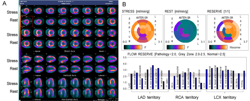
Fig. 4 Post-PCI 13N-ammonia PET findings. (A) Markedly improved myocardial perfusion over entire left ventricle was noted, (B) Coronary flow reserve was improved in left anterior descending artery and left circumflex artery.
Reference
-
1. Pijls NH, Fearon WF, Tonino PA, Siebert U, Ikeno F, Bornschein B, et al. Fractional flow reserve versus angiography for guiding percutaneous coronary intervention in patients with multivessel coronary artery disease: 2-year follow-up of the FAME (Fractional Flow Reserve Versus Angiography for Multivessel Evaluation) study. J Am Coll Cardiol. 2010; 56:177–184.
Article2. Nam CW, Mangiacapra F, Entjes R, Chung IS, Sels JW, Tonino PA, et al. Functional SYNTAX score for risk assessment in multivessel coronary artery disease. J Am Coll Cardiol. 2011; 58:1211–1218.
Article3. Gould KL, Johnson NP, Bateman TM, Beanlands RS, Bengel FM, Bober R, et al. Anatomic versus physiologic assessment of coronary artery disease. Role of coronary flow reserve, fractional flow reserve, and positron emission tomography imaging in revascularization decision-making. J Am Coll Cardiol. 2013; 62:1639–1653.4. Pijls NH, De Bruyne B, Peels K, Van Der Voort PH, Bonnier HJ, Bartunek J, Koolen JJ, et al. Measurement of fractional flow reserve to assess the functional severity of coronaryartery stenoses. N Engl J Med. 1996; 334:1703–1708.
Article5. Johnson NP, Kirkeeide RL, Gould KL. Is discordance of coronary flow reserve and fractional flow reserve due to methodology or clinically relevant coronary pathophysiology? JACC Cardiovasc Imaging. 2012; 5:193–202.
Article6. Yoshinaga K, Chow BJ, Williams K, Chen L, deKemp RA, Garrard L, et al. What is the prognostic value of myocardial perfusion imaging using rubidium-82 positron emission tomography? J Am Coll Cardiol. 2006; 48:1029–1039.
Article7. Herzog BA, Husmann L, Valenta I, Gaemperli O, Siegrist PT, Tay FM, et al. Long-term prognostic value of 13N-ammonia myocardial perfusion positron emission tomography added value of coronary flow reserve. J Am Coll Cardiol. 2009; 54:150–156.
Article8. Ziadi MC, Dekemp RA, Williams KA, Guo A, Chow BJ, Renaud JM, et al. Impaired myocardial flow reserve on rubidium-82 positron emission tomography imaging predicts adverse outcomes in patients assessed for myocardial ischemia. J Am Coll Cardiol. 2011; 58:740–748.
Article9. Fiechter M, Gebhard C, Ghadri JR, Fuchs TA, Pazhenkottil AP, Nkoulou RN, et al. Myocardial perfusion imaging with 13N-ammonia PET is a strong predictor for outcome. Int J Cardiol. 2013; 167:1023–1026.
Article10. Tonino PA, Fearon WF, De Bruyne B, Oldroyd KG, Leesar MA, Ver Lee PN, et al. Angiographic versus functional severity of coronary artery stenoses in the FAME study fractional flow reserve versus angiography in multivessel evaluation. J Am Coll Cardiol. 2010; 55:2816–2821.
Article
- Full Text Links
-
- Actions
-
Cited
- CITED
-
- Close
- Share
-
- Similar articles
-
- Evaluation of Myocardial Blood Flow and Coronary Flow Reserve Using Positron Emission Tomography
- Percutaneous coronary intervention in patients with multi-vessel coronary artery disease: a focus on physiology
- Physiologic Assessment after Coronary Stent Implantation
- Indirect Radionuclide Coronary Angiography to Evaluate Gradients of Myocardial Blood Flow and Flow Reserve Through Coronary Stenosis Using N-13 Ammonia PET/CT
- Physiologic Assessment of Coronary Artery Disease: Past, Present and Future
